

Juvenile xanthogranuloma involving concurrent iris and skin: Clinical, pathological and molecular pathological evaluations
- PMID: 29468209
- PMCID: PMC5787822
- DOI: 10.1016/j.ajoc.2017.09.004
Juvenile xanthogranuloma involving concurrent iris and skin: Clinical, pathological and molecular pathological evaluations
Abstract
Purpose: To report a case of juvenile xanthogranuloma involving the iris and skin that clincally was diagnosed with an obvious cutaneous lesion.
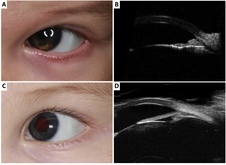
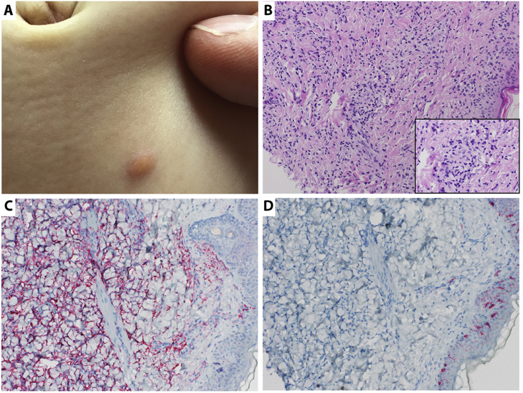
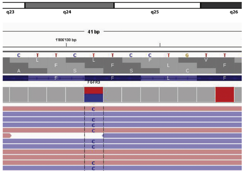
Observations: A four month-old girl with hyphema and increased intraocular pressure of the left eye persisting for 2 weeks. A suspicious yellow-brown mass with nodular surface and traversed by irregular vascularization was noted on the inferior iris surface. Ultrasound biomicroscopy (UBM; 35 MHz) of the mass revealed multiple nodular irregular hyperreflective lesions in the peripheral iris. Using a biopsy of an obvious cutaneous abdominal skin lesion a diagnosis was made based on histopathological analyses. The biopsy showed dense dermal infiltrate consisting of foamy histiocytes. Additional stains revealed CD68 positivity and CD1a and S100 negativity. This mass revealed histopathologic features identical to juvenile xanthogranuloma and was concurrent with the iris lesion. Next-generation sequencing using Ion AmpliSeqTM Cancer Hotspot Panel revealed a missense mutation of FGFR3 (p.F386L).
Conclusion and importance: The diagnosis of a xanthogranuloma of the iris with hyphema can be made easier in patients with obvious cutaneous lesions as described in our case. The significance of FGFR3 mutation in association with JXG is unknown and should be further investigated.
Keywords: Cutaneous lesion; FGFR3 mutation; Iris; Juvenile xanthoganuloma; Ultrasound biomicroscopy.
Figures
References
-
- Chang M.W. Update on juvenile xanthogranuloma: unusual cutaneous and systemic variants. Semin Cutan Med Surg. 1999;18(3):195–205. - PubMed
-
- Danzig C.J., Shields C.L., Mashayekhi A., Ehya H., Manquez M.E., Shields J.A. Fluorescein angiography of iris juvenile xanthogranuloma. J Pediatr Ophthalmol Strabismus. 2008;45(2):110–112. - PubMed
-
- Hernandez-Martin A., Baselga E., Drolet B.A., Esterly N.B. Juvenile xanthogranuloma. J Am Acad Dermatol. 1997;36(3 Pt 1):355–367. quiz 368–359. - PubMed
-
- Samara W.A., Khoo C.T., Say E.A. Juvenile xanthogranuloma involving the eye and ocular adnexa: tumor control, visual outcomes, and globe salvage in 30 patients. Ophthalmology. 2015;122(10):2130–2138. - PubMed
Publication types
LinkOut - more resources
Full Text Sources
Other Literature Sources
