Review
. 2018 May;48(5):735-744.
doi: 10.1007/s00247-018-4093-0. Epub 2018 Feb 21.

Testicular torsion with preserved flow: key sonographic features and value-added approach to diagnosis

Affiliations
Review

Testicular torsion with preserved flow: key sonographic features and value-added approach to diagnosis

Anjum N Bandarkar et al. Pediatr Radiol. 2018 May.

Abstract

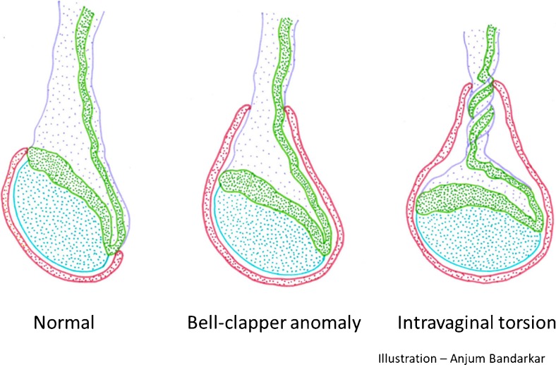
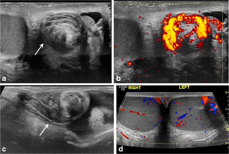
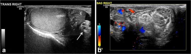
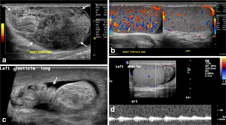
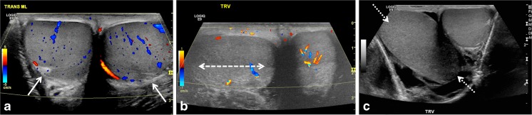
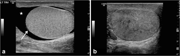
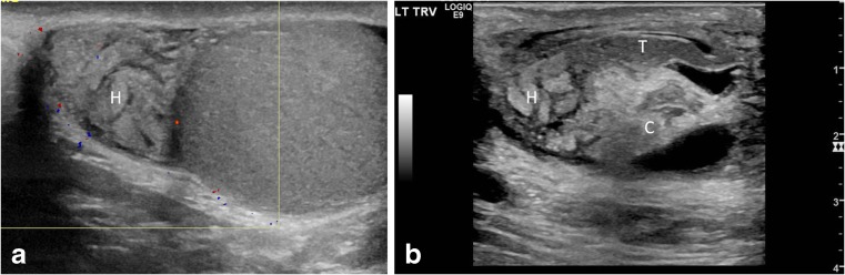
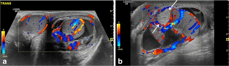
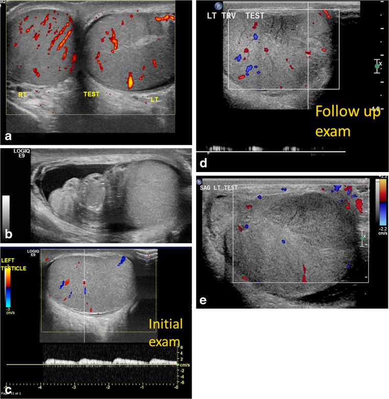
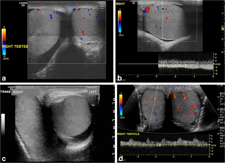
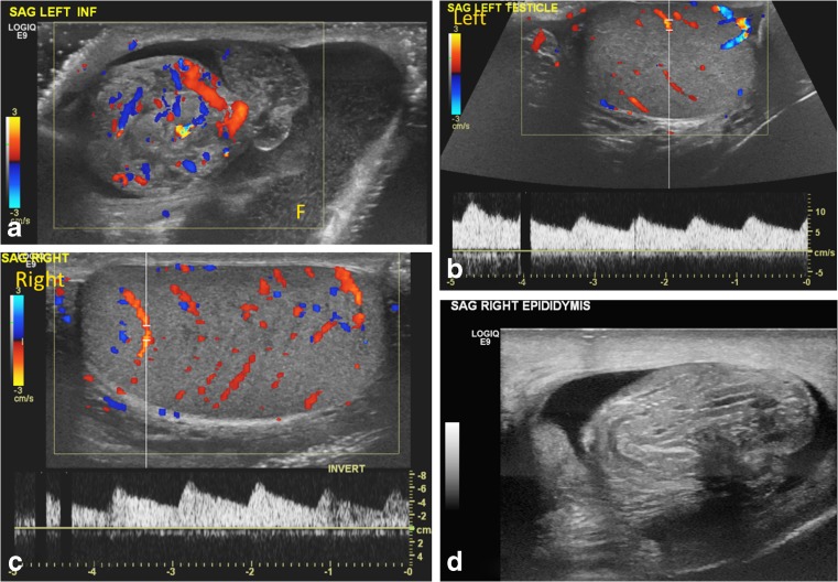
Testicular sonography has contributed greatly to the preoperative diagnosis of testicular torsion in the pediatric patient and is the mainstay for evaluation of acute scrotal pain. Despite its high sensitivity and specificity, both false-negative and false-positive findings occur. Presence of documented Doppler flow within the testis might be a dissuading factor for surgical exploration with resultant testicular loss in the false-negative cases. Our goal is to illustrate key sonographic features in the spectrum of testicular torsion with preserved testicular flow, and to describe how to differentiate testicular torsion from epididymitis in order to avoid the under-diagnosis of testicular torsion. We simplify the anatomy of the bell clapper testis. We also describe our sonographic protocol for testicular torsion and share valuable tips from our approach to challenging cases.

Keywords: Bell clapper anomaly; Children; Epididymis; Scrotum; Testis; Torsion; Ultrasound; Whirlpool sign.


Conflict of interest statement

None

Figures

Fig. 1
Fig. 1
Anatomy of the normal testis, bell clapper anomaly and intravaginal testicular torsion. Blue testis, Green epididymis, Lavender spermatic cord and vessels, Red tunica vaginalis
Fig. 2
Fig. 2
“Whirlpool sign” of spermatic cord. a Gray-scale transverse US image of upper left scrotal sac shows an eddy swirl (arrow) of the spermatic cord suggesting torsion of the cord. This 12-year-old boy woke with acute left testicular pain and experienced nausea and vomiting along with the pain. b Power Doppler US image of the same twisted cord shows concentric pattern of preserved flow in the vessels of the twisted cord. The flow in the left testis (not shown) was minimally decreased compared to the right side and bilateral bell clapper deformity was found during orchiopexy along with complete torsion of the left testis with 360° twist. c Gray-scale longitudinal US image of the left scrotum in a 13-year-old boy with 1 day of left-side pain shows abrupt spiral twisting of the spermatic cord (arrow) at the external inguinal ring, creating a whirlpool sign. d Color Doppler transverse image of the testes in the same boy as in (c) shows preserved and symmetrical flow bilaterally. After manual detorsion in the emergency room, he underwent orchiopexy and was diagnosed with intermittent torsion
Fig. 3
Fig. 3
Intermittent torsion in a 17-year-old boy who presented with 5 h of acute right testicular pain after a game of football. He had experienced 6–7 similar episodes in the last 2 years where the pain had spontaneously resolved. Cremasteric reflex was absent on the right. a Gray-scale transverse US image of the right testis shows a redundant spermatic cord (arrow) occupying the medial half of the scrotal sac, with a mildly edematous epididymis (E) adjacent to it. The echogenic mediastinum testis faced medially instead of posterolaterally, which was concerning for altered testicular lie. b Color Doppler longitudinal image of the right scrotum shows excess and tortuous spermatic cord bunched up in the scrotal sac superior to the testis and formation of pseudomass, suggesting torsion of the spermatic cord. Note that this extratesticular pseudomass is not hyperemic and should not be confused with epididymitis. Orchiopexy was recommended; however the family chose to wait because his pain improved. Elective orchiopexy was performed 7 months later and bilateral bell clapper anomaly was noted; he was diagnosed with intermittent torsion
Fig. 4
Fig. 4
Redundant spermatic cord and enlarged epididymal-cord complex in the setting of preserved testicular flow. a Power Doppler US image in a 15-year-old boy who presented with acute right testicular pain shows an avascular pseudomass (arrows) along the superior aspect of the testis. b Scrotal color Doppler sonography shows preserved and increased testicular flow from manual detorsion but there is a bunched up cord as seen in Fig 4a, forming an enlarged epididymal-cord complex or extratesticular pseudomass. This suggests that the cord was tangled with and inseparable from the epididymis, with the point of twist forming the torsion knot. c Gray-scale US image at the level of the external inguinal ring in a 12-year-old boy with 24 h of acute left testicular pain shows a twisted redundant cord (arrow) in the lower inguinal canal (identified retrospectively) with a small surrounding hydrocele. d Color and pulsed wave Doppler US in the same boy as in (c) shows preserved left testicular flow. Because of the long duration of pain, which had begun to improve at the time of exam, and presence of intratesticular flow, he was given a diagnosis of epididymitis. He returned a week later with an infarcted left testis that showed ultrasound findings of late torsion. This example reiterates that duration of pain and presence of intratesticular flow should not deter one from looking for the twisted cord because the latter can clinch the diagnosis
Fig. 5
Fig. 5
Testicular lie — normal and abnormal. a Color Doppler transverse US image of the testes in a 12-year-old boy with mild groin pain demonstrates normal vertical lie with the testes seen in round cross-sections and with the mediastinum testis (arrows) directed posterolaterally. b Color Doppler transverse US image of both testes in a 14-year-old boy who woke with acute right scrotal pain demonstrates abnormal horizontal lie of the right testis (arrow) with slightly decreased intratesticular flow compared to the normal left side. He had experienced similar episodes of pain in the past and was diagnosed with intermittent torsion. During orchiopexy 12 h later, a bell clapper deformity was noted bilaterally. c Gray-scale transverse US image of both testes in a 16-year-old boy with right testicular pain demonstrates abnormal oblique lie of the right testis (arrows), which is oriented diagonally compared to the normal left side. Intermittent torsion was diagnosed intraoperatively
Fig. 6
Fig. 6
Globular testicular enlargement and heterogeneous echotexture. a Gray-scale transverse US image of the left scrotum in a 13-year-old boy with 3 days of intermittent left testicular pain shows globular shape of left testis with horizontal lie (arrow), decreased flow and redundant spermatic cord (neither shown), and reactive hydrocele (star). He was found to have a 180° twist on the left with abnormal epididymal attachment (the epididymis was attached only to the superior pole, suggesting bell clapper deformity) and he underwent bilateral orchiopexy after left detorsion. b Gray-scale longitudinal US image of the right testis in a 16-year-old boy with acute right testicular pain shows heterogeneous echotexture of the right testis; decreased flow and redundant spermatic cord were also present (not shown). Flow returned after detorsion and bilateral orchiopexy was performed. Intermittent torsion was diagnosed intraoperatively
Fig. 7
Fig. 7
Epididymal enlargement without hyperemia. a Color Doppler longitudinal US image of the epididymal head and testis in a 12-year-old boy who woke with acute left testicular pain and nausea. Image shows an enlarged epididymal head (H) with a swollen lobular appearance and lack of intrinsic color flow. The intratesticular flow was decreased. He underwent emergent bilateral orchiopexy for a complete left torsion, and a bell clapper deformity was appreciated. Even though the epididymis was enlarged, it was avascular, thus ruling out epididymitis. b Gray-scale longitudinal US image of the epididymal-cord complex in a 7-year-old boy with 3 days of intermittent left testicular pain shows an edematous epididymal head (H) with a tangled echogenic cord (C) while the epididymal tail (T) appears relatively uninvolved. His left testis, however, showed somewhat decreased flow and he was diagnosed with partial torsion. A 360° twist was found during orchiopexy along with bilateral classic bell clapper deformity
Fig. 8
Fig. 8
Epididymitis and testicular appendage torsion. a Transverse color Doppler US image of both testes in a 9-year-old boy with left-side pain shows hyperemia in the left testis and thickened epididymis (E) with surrounding hydrocele and left scrotal wall edema, all favoring left epididymo-orchitis. b Longitudinal color Doppler US image of the left scrotum in a 10-year-old boy with acute pain shows a globular, enlarged, echogenic and avascular nodular structure (arrows) at the superior pole of the testis, compatible with torsion of the testicular appendage. Not infrequently, reactive inflammatory change involves the ipsilateral testis and epididymis
Fig. 9
Fig. 9
Partial torsion in a 13-year-old boy who presented with a 2-week history of intermittent left testicular pain and swelling. a Transverse power Doppler US image of both testes shows bilateral preserved flow with a slightly globular-appearing left testis. b Gray-scale longitudinal US image of the left testis and epididymis shows an enlarged left epididymal-cord complex and surrounding small hydrocele. The boy was discharged with a diagnosis of epididymitis because of the long confounding duration of symptoms and presence of intratesticular flow. c Color and pulsed Doppler US image of the left testis at initial presentation shows preserved intrinsic flow with normal arterial waveform. d Follow-up exam after 3 weeks demonstrates abnormally dampened left testicular flow and waveforms. e Color Doppler US image of left testis during at follow-up for persistent pain shows interval significantly enlarged left testis with decreased vascularity and a focal hypovascular parenchymal area concerning for infarct in the setting of incomplete torsion. He underwent left detorsion with bilateral orchiopexy
Fig. 10
Fig. 10
Complete torsion with preserved flow. a Transverse color Doppler US image of both testes in a 13-year-old boy with 8 h of right-side acute pain shows oblique lie and globular enlargement of the right testis with preserved flow and surrounding small reactive hydrocele. There is a linear transducer artifact at mid-testis level. b Longitudinal color Doppler US image of the right testis shows a globular testis with intratesticular flow. During surgery, complete torsion was diagnosed with a 360° twist and bilateral bell clapper deformity. c Transverse gray-scale image of both testes in a 15-year-old boy with acute right testicular pain shows horizontal lie of the right testis. The asymptomatic left testis incidentally demonstrates a medially directed mediastinum testis and trace fluid in the tunica vaginalis. d Transverse color Doppler US image demonstrates preserved intratesticular flow thought to be symmetrical. Complete torsion of the right testis was diagnosed at surgery, and bilateral orchiopexy was performed post detorsion
Fig. 11
Fig. 11
Bilateral partial torsion in a 15-year-old boy who presented with acute left testicular pain. a Color Doppler US image of the upper left scrotal sac shows an enlarged epididymal-cord complex with flow within, and surrounding complex hydrocele with mobile echoes. b, c Preserved slightly asymmetrical — left (b) less than right (c) — intratesticular flow and symmetrical waveforms are appreciated. The enlarged epididymal-cord complex was confused for epididymitis. d Follow-up exam after 4 months for recurrent testicular pain, now on the right side, shows an enlarged epididymal-cord complex in the right scrotum. Bilateral partial torsion (270° twist on the right side and 90° twist on the left side) and bilateral bell clapper anomaly were detected at orchiopexy

References

    1. Munden M, Williams J, Zhang W, et al. Intermittent testicular torsion in the pediatric patient: sonographic indicators of a difficult diagnosis. AJR Am J Roentgenol. 2013;201:912–918. doi: 10.2214/AJR.12.9448. - DOI - PubMed
    1. Murphy F, Fletcher L, Pease P. Early scrotal exploration in all cases is the investigation and intervention of choice in all cases of acute pediatric scrotum. Pediatr Surg Int. 2006;2:413–416. doi: 10.1007/s00383-006-1681-0. - DOI - PubMed
    1. Vikram S, Bhatt S, Rubens D. Sonographic evaluation of testicular torsion. Ultrasound Clinics. 2006;1:55–66. doi: 10.1016/j.cult.2005.09.006. - DOI
    1. Martin A, Rushton H. The prevalence of bell clapper anomaly in the solitary testis in cases of prior perinatal torsion. J Urol. 2014;191:1573–1577. doi: 10.1016/j.juro.2013.09.013. - DOI - PubMed
    1. Caesar R, Kaplan G. Incidence of the bell-clapper deformity in an autopsy series. Urology. 1994;44:114–116. doi: 10.1016/S0090-4295(94)80020-0. - DOI - PubMed

LinkOut - more resources