. 2018 Jan-Dec:23:2156587218757649.
doi: 10.1177/2156587218757649.

Protective Effect of Kolaviron on Cyclophosphamide-Induced Cardiac Toxicity in Rats

Affiliations

Protective Effect of Kolaviron on Cyclophosphamide-Induced Cardiac Toxicity in Rats

Joseph Gbenga Omole et al. J Evid Based Integr Med. 2018 Jan-Dec.

Abstract

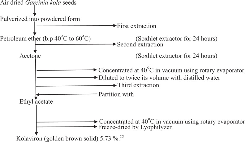
Background: Cyclophosphamide (CP) is a nitrogen mustard alkylating drug used for the treatment of chronic and acute malignant lymphomas, myeloma, leukemia, neuroblastoma, adenocarcinoma, retinoblastoma, breast carcinoma, and immunosuppressive therapy. Despite its vast therapeutic uses, it is known to cause severe cardiac toxicity. Kolaviron (KV), a Garcinia kola seed extract containing a mixture of flavonoids, is reputed for its antioxidant and membrane stabilizing properties.

Objective: This study investigated the protective effect of KV on CP-induced cardiotoxicity in rats.

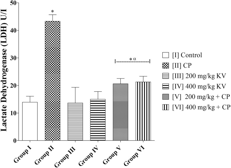
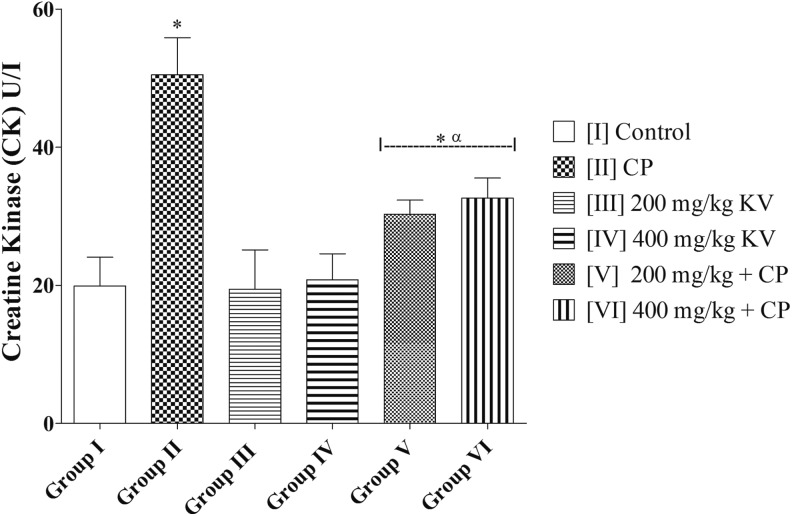
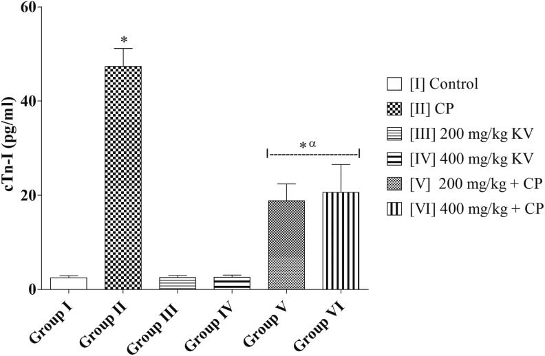
Methods: Thirty rats were used, and they were divided into 6 groups of 5 rats each. Group I received 2 mL/kg propylene glycol orally for 14 days; group II received CP (50 mg/kg/d, intraperitoneally [i.p.]) for 3 days; groups III and IV received 200 and 400 mg/kg/d KV, respectively, orally for 14 days and groups V and VI were pretreated with 200 and 400 mg/kg/d KV, respectively, orally for 14 days followed by CP (50 mg/kg/d, i.p.) for 3 days.

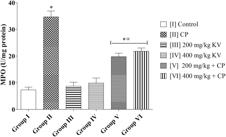
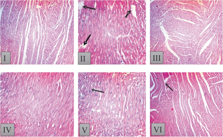
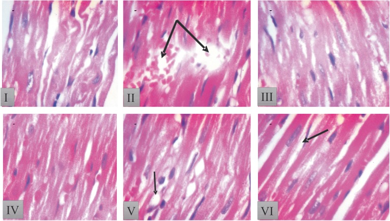
Results: CP treatment resulted in a significantly lower food consumption and body weight in rats. The lactate dehydrogenase and creatine kinase enzymes in cardiac tissues of rats treated with CP were significantly higher. In cardiac tissues, 3-day doses of CP resulted in significantly higher heart weight, cardiac troponin I, myeloperoxidase, malondialdehyde, hydrogen peroxide and lower superoxide dismutase, catalase, glutathione peroxidase activities, and reduced glutathione levels. Histological examination of cardiac tissues showed sign of necrosis of myocardium after CP treatment. However, administration of KV at 200 and 400 mg/kg for 14 days prior to CP treatment, increase food consumption, body weight, and attenuates the biochemical and histological changes induced by CP.

Conclusions: These results revealed that KV attenuates CP-induced cardiotoxicity by inhibiting oxidative stress and preserving the activity of antioxidant enzymes.

Keywords: antioxidant; cardiac troponin I; cardiotoxicity; cyclophosphamide; kolaviron; oxidative stress.


Conflict of interest statement

Declaration of Conflicting Interests: The authors declared no potential conflicts of interest with respect to the research, authorship, and/or publication of this article.

Figures

None
Scheme illustration of extraction process of kolaviron.
Figure 1.
Figure 1.
Effects of kolaviron on lactate dehydrogenase (LDH) activities in rats treated with cyclophosphamide (CP). Values are given as mean ± standard error of the mean (SEM) (n = 5). *P < .05 compared with control and α P < .05 compared with CP.
Figure 2.
Figure 2.
Effects of kolaviron on creatine kinase (CK) activities in rats treated with cyclophosphamide (CP). Values are given as mean ± standard error of the mean (SEM) (n = 5). *P < .05 compared with control and α P < .05 compared with CP.
Figure 3.
Figure 3.
Effects of kolaviron on myeloperoxidase (MPO) activities in rats treated with cyclophosphamide (CP). Values are given as mean ± standard error of the mean (SEM) (n = 5). *P < .05 compared with control and α P < .05 compared with CP.
Figure 4.
Figure 4.
Effects of kolaviron on cardiac troponin I (cTn I) in rats treated with cyclophosphamide (CP). Values are given as mean ± standard error of the mean (SEM) (n = 5). *P < .05 compared with control and α P < .05 compared with CP.
Figure 5.
Figure 5.
Histopathological result of heart tissue in [I] control: showed normal histoarchitecture of the heart with no visible lesion; [II] Cyclophosphamide (CP) treated. CP-treated group [II] shows distorted and wavy myocardial fibers with focal fatty change in some area of myocardial fibers. Loss of cellular constituents of the myocardial cells (black arrow), myofibrillar loss and hypertrophic myocardial fiber with inflammation; [III] 200 mg/kg kolaviron (KV) showed intact myocardial fibers, but focal areas of hyalinization and some wavy myocardial fibers; [IV] 400 mg/kg KV showed a few wavy myocardial fibers as well as few losses of cellular constituents; [V] 200 mg/kg KV + CP treated: showed improvement in histoarchitecture with mild waviness of myofibers as well as separation of some myocardial fibers and [VI] 400 mg/kg KV + CP treated rats; showed mild fatty change in some muscle cells and separation of some myocardial fibers. Sections of heart tissues of rats were stained with hematoxylin-eosin (magnification 100×).
Figure 6.
Figure 6.
Histopathological result of heart tissue in [I] control: rats heart tissue showed intact myocardial fibers and pericardium; [II] cyclophosphamide (CP) alone: rats heart tissue produced massive change in the myocardium showing a varying degree of vacuolar changes in the cardiac muscle fibers mainly in the form of degeneration of myocardial fibers, vacuolization of the cardiomyocytes, infiltration of inflammatory cells, myofibrillar loss, and hypertrophic myocardial fiber with inflammation (double arrow); [III] 200 mg/kg kolaviron (KV) showed intact myocardial fibers, but focal areas of hyalinization and some wavy myocardial fibers; [IV] 400 mg/kg KV showed a few wavy myocardial fibers as well as few losses of cellular constituents; [V] 200 mg/kg KV + CP treated showed improvement in histoarchitecture with mild waviness of myofibers as well as separation of some myocardial fibers; and [VI] 400 mg/kg KV + CP treated rats showed mild fatty change in some muscle cells and separation of some myocardial fibers. Sections of heart tissues of rats were stained with hematoxylin-eosin (magnification 400×).

References

    1. Demirer T, Buckner CD, Appelbaum FR, et al. Busulfan, cyclophosphamide and fractionated total body irradiation for autologous or syngeneic marrow transplantation for acute and chronic myelogenous leukemia: phase I dose escalation of busulfan based on targeted plasma levels. Bone Marrow Transplant. 1996;17:491–495. - PubMed
    1. Itescu SE, Burke K, Lietz R, et al. Intravenous pulse administration of cyclophosphamide is an effective and safe treatment for sensitized cardiac allograft recipients. Circulation. 2002;105:1214–1219. - PubMed
    1. Pagel JM, Appelbaunm FR, Earyis IF, et al. 131I-anti-CD45 antibody plus busulfan and cyclophosphamide before allogeneic hematopoietic cell transplantation for treatment of acute myeloid leukemia in first remission. Blood. 2006;107:2184–2191. - PMC - PubMed
    1. Zhang J, Tian Q, Zhou SF. Clinical pharmacology of cyclophosphamide and ifosfamide. Curr Drug Ther. 2006;1:55-84(30).
    1. Hirano A, Shimizu T, Watanabe O, et al. Epirubicin and cyclophosphamide followed by docetaxel as primary systemic chemotherapy in locally advanced breast cancer. Anticancer Res. 2008;28:4137–4142. - PubMed

MeSH terms

LinkOut - more resources