. 2018;9(5):400-421.
doi: 10.1080/19490976.2018.1441664. Epub 2018 May 31.

Prenatal androgen exposure causes hypertension and gut microbiota dysbiosis

Affiliations

Prenatal androgen exposure causes hypertension and gut microbiota dysbiosis

Shermel B Sherman et al. Gut Microbes. 2018.

Abstract

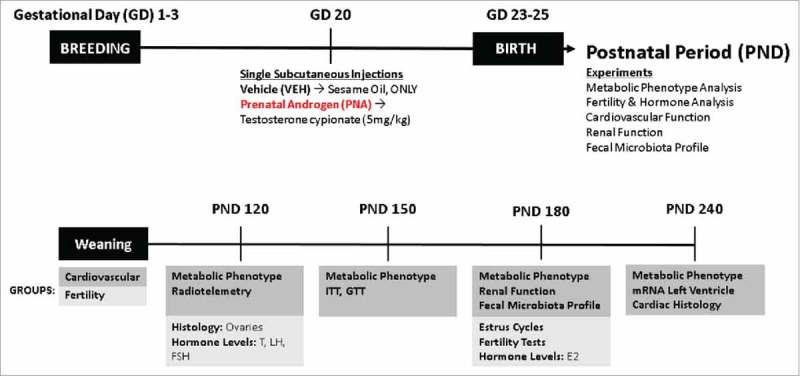
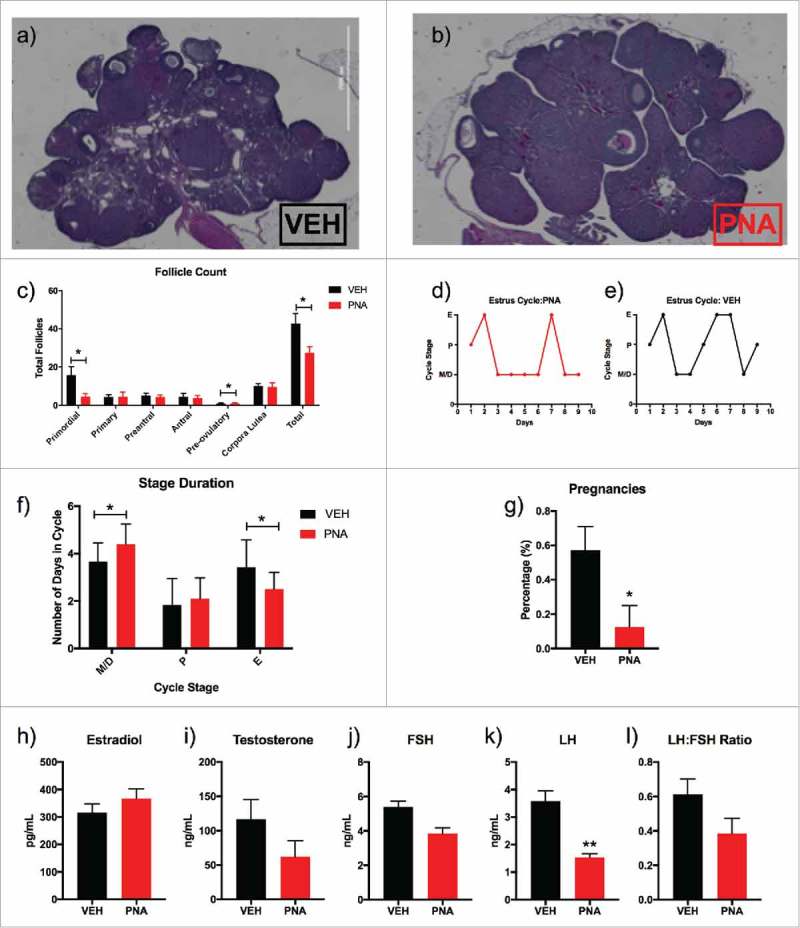
Background: Conditions of excess androgen in women, such as polycystic ovary syndrome (PCOS), often exhibit intergenerational transmission. One way in which the risk for PCOS may be increased in daughters of affected women is through exposure to elevated androgens in utero. Hyperandrogenemic conditions have serious health consequences, including increased risk for hypertension and cardiovascular disease. Recently, gut dysbiosis has been found to induce hypertension in rats, such that blood pressure can be normalized through fecal microbial transplant. Therefore, we hypothesized that the hypertension seen in PCOS has early origins in gut dysbiosis caused by in utero exposure to excess androgen. We investigated this hypothesis with a model of prenatal androgen (PNA) exposure and maternal hyperandrogenemia by single-injection of testosterone cypionate or sesame oil vehicle (VEH) to pregnant dams in late gestation. We then completed a gut microbiota and cardiometabolic profile of the adult female offspring.

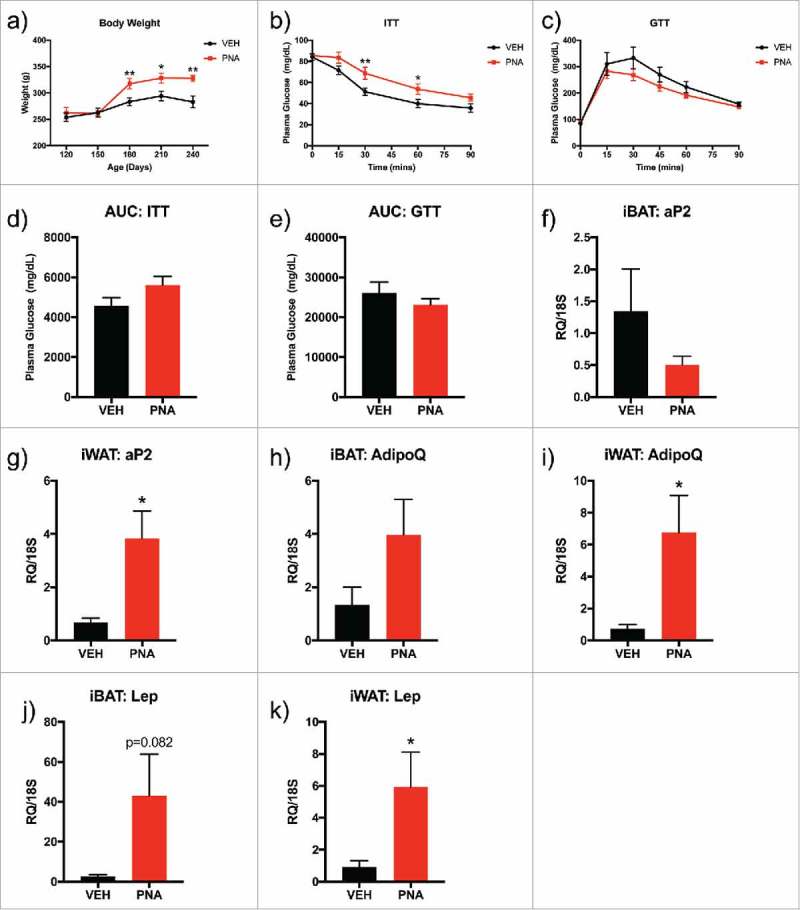
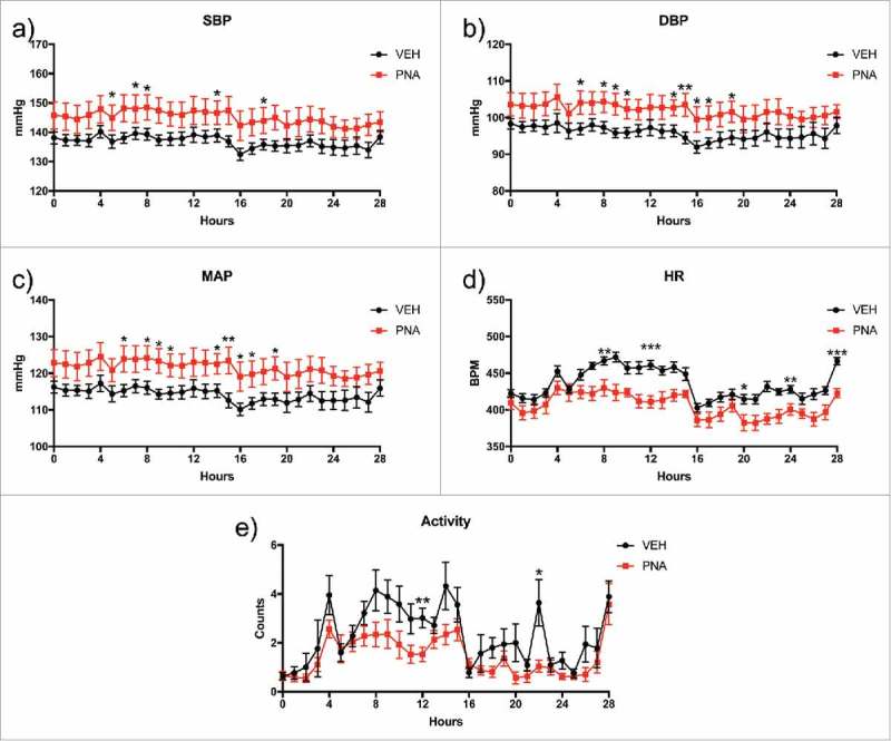
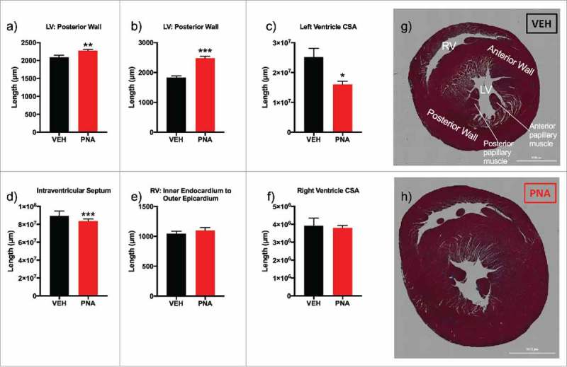
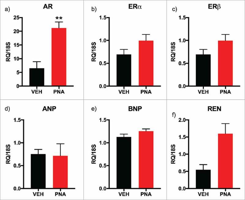
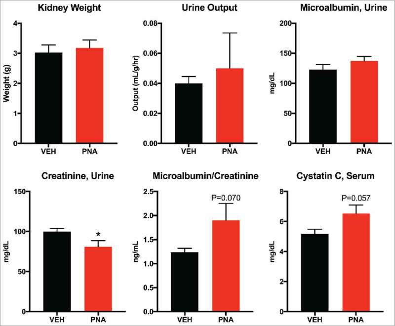
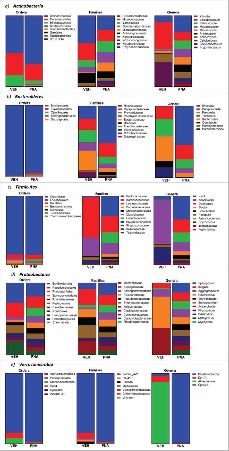
Results: The metabolic assessment revealed that adult PNA rats had increased body weight and increased mRNA expression of adipokines: adipocyte binding protein 2, adiponectin, and leptin in inguinal white adipose tissue. Radiotelemetry analysis revealed hypertension with decreased heart rate in PNA animals. The fecal microbiota profile of PNA animals contained higher relative abundance of bacteria associated with steroid hormone synthesis, Nocardiaceae and Clostridiaceae, and lower abundance of Akkermansia, Bacteroides, Lactobacillus, Clostridium. The PNA animals also had an increased relative abundance of bacteria associated with biosynthesis and elongation of unsaturated short chain fatty acids (SCFAs).

Conclusions: We found that prenatal exposure to excess androgen negatively impacted cardiovascular function by increasing systolic and diastolic blood pressure and decreasing heart rate. Prenatal androgen was also associated with gut microbial dysbiosis and altered abundance of bacteria involved in metabolite production of short chain fatty acids. These results suggest that early-life exposure to hyperandrogenemia in daughters of women with PCOS may lead to long-term alterations in gut microbiota and cardiometabolic function.

Keywords: Microbiota; PCOS; androgens; blood pressure; cardiometabolic; cardiovascular disease; cystatin C; hyperandrogenemia; hypertension; kidney; testosterone.


Figures

Figure 1.
Figure 1.
Schematic of Experimental Design and Animal Timeline. The animals were separated into two experimental cohorts: cardiovascular and fertility. The cardiovascular cohort experiments were performed from PND 120 to PND 240. Metabolic phenotype includes monthly body weight measurements, fecal sample collection at PND 120 (VEH = 10, PNA = 10), insulin tolerance tests (ITT), and glucose tolerance tests (GTT) (VEH = 12, PNA = 10). Cardiovascular function included Radiotelemetry performed at PND 120 (VEH = 12, PNA = 10), mRNA of the left ventricle of the heart at PND 240 and cardiac histology for assessment of cardiac hypertrophy (VEH = 4, PNA = 4). Renal function included serum and urine assays performed at PND 180 (VEH = 10, PNA = 10). The fertility cohort experiments were performed at PND 120 and PND 180. These experiments included ovarian histology at PND 120 and serum hormone analysis of testosterone (T), luteinizing hormone (LH), and follicle-stimulating hormone (FSH) (VEH = 12, PNA = 10). At PND 180, serum was collected for hormone levels of estradiol (E2), vaginal cytology was collected for assessment of estrus cycles (VEH = 12, PNA = 10). Finally, fertility tests were performed to determine pregnancy rates (VEH = 14, PNA = 8).
Figure 2.
Figure 2.
Metabolic Assessment: Body Composition, Glucose and Insulin Tolerance Tests, mRNA Expression of Adipokines within Adipose Tissue. (a) Body weight was measured from PND 120 to PND 240. (b) Glucose tolerance test (GTT) was performed at PND150 with intraperitoneal injection (IP) of dextrose and plasma glucose levels were recorded for time 0 to 120 minutes. (c) Insulin tolerance test (ITT) was performed at PND150 with IP injection of insulin and plasma glucose levels were recorded for time 0 to 90 minutes. (d) Area under the curve (AUC) for GTT was calculated for all plasma glucose levels between 0–120 minutes (e) Area under the curve (AUC) for ITT was calculated for all plasma glucose levels between 0–90 minutes. Bars represent mean and lines represent standard error (SEM); (f) iBAT aP2 mRNA expression, g) iWAT aP2 mRNA expression, (h) ibAT AdipoQ mRNA expression, (i) iWAT AdipoQ mRNA expression, (j) iBAT Lep mRNA expression, (k) iWAT Lep mRNA expression. The tissue was dissected from sacrificed animals at PND120 and the sample size was n = 6–7 animals per group (VEH or PNA). Bars represent means and the lines represent the standard error of the mean (SEM). Symbols in either black (VEH) or red (PNA) represent the distribution of relative expression (RQ) values within each group. All genes were normalized to the RQ of 18S.
Figure 3.
Figure 3.
Fertility and Serum Hormone Analysis. (a) Hematoxylin and eosin stained ovary of female vehicle rat at PND 120; (b) Hematoxylin and eosin stained ovary of female PNA rat at PND 120; (c) Total follicle count of primordial, primary, preantral, antral, preovulatory, corpora lutea, and total follicle count; (d) Estrous cycle data for nine consecutive days of vaginal smearing of vehicle (n = 12) rats; (e) Estrous cycle data for nine consecutive days of vaginal smearing of and prenatal androgen rats (n = 11); (f) Duration of estrus cycle and number of days spent in each stage for PNA animals compared to vehicle; (g) Pregnancy success rates for vehicle (n = 14) and PNA (n = 8) rats at PND 180; (h) Serum beta-estradiol levels for VEH and PNA animals measured in picograms per milliliter at PND 180; (i) Serum testosterone levels for VEH and PNA animals measured in nanograms per milliliter at PND 120; (j) Serum follicle stimulating hormone (FSH) levels for VEH and PNA animals measured in nanograms per milliliter in animals at PND 180; (k) Serum luteinizing hormone (LH) levels for VEH and PNA animals measured in nanograms per milliliter in animals at PND 180; and (l) LH:FSH ratio measured for animals at PND 180.
Figure 4.
Figure 4.
Radiotelemetry Blood Pressure, Heart Rate, and Locomotor Activity. Radiotelemetry analysis was performed for moving averages every hour for 28 hours with standard error calculated for each hour. The shaded areas on the graph represent night hours. (a) Systolic blood pressure (SBP), (b) diastolic blood pressure (DBP), (c) Mean arterial pressure (MAP), (d) heart rate (BPM), and (e) locomotor activity.
Figure 5.
Figure 5.
Cardiac morphology and gross anatomical measurements. (a) left ventricle (LV) anterior muscle wall; (b) LV posterior muscle wall; (c) left ventricle cross sectional area (CSA); (d) intraventricular septum (e) right ventricle (RV) inner endocardium to outer epicardium measurement; (f) right ventricle cross sectional area (CSA); (g) Masson's trichrome stain of short-axis cut with angulation to expose left ventricle papillary muscles and apex of right ventricle (RV) of VEH female rat; (h) Masson's trichrome stain of short-axis cut of heart with angulation to expose left ventricle papillary muscles and apex of right ventricle of PNA female rat.
Figure 6.
Figure 6.
mRNA Expression of Sex Steroid Receptors, Cardiac Hypertrophy Markers, and Renin within Left Ventricle (LV) of the Heart. (a) AR, androgen receptor mRNA expression; (b) ERα, estrogen receptor alpha mRNA expression; (c) ERβ, estrogen receptor beta mRNA expression; (d) ANP, atrial naturetic protein mRNA expression; (e) BNP, B-type (brain) naturetic protein mRNA expression; (f) Ren, renin mRNA expression. All genes were normalized to 18S expression. All bars represent the mean values of relative expression (RQ) and lines represent standard error of the mean.
Figure 7.
Figure 7.
Renal Function Assessment of Serum Cystatin C and Urine Microalbumin and Creatinine. (a) paired kidney weight was measured immediately after dissection. (b) urine output was calculated from 24-hour urine volume in milliliters divided by body weight in grams, divided by 24-hours. (c) urine creatinine was measured from 24-hour fasting urine. (d) microalbumin was measured from 24-hour fasting urine. (e) microalbumin/creatinine ratio. (f) cystatin C concentration was measured using serum collected from PND180 animals.
Figure 8.
Figure 8.
Relative abundance of bacteria within the most abundance phylum: Actinobacteria, Bacteroidetes, Firmicutes, Proteobacteria, and Verrucomicrobia. Relative abundance of bacteria at the phylum, order, family and genera levels were determined using operational taxonomic units for each. The graphs represent the percentages for the relative abundance of bacteria from the total OTU of each phylum, order, family, or genera.

Similar articles

Cited by

References

    1. Perez-Sepulveda A, Monteiro LJ, Dobierzewska A, España-Perrot PP, Venegas-Araneda P, Guzmán-Rojas AM, González Mí I, Palominos-Rivera M, Irarrazabal CE, Figueroa-Diesel H, et al.. Placental Aromatase Is Deficient in Placental Ischemia and Preeclampsia. PLoS One. 2015;10. doi:10.1371/journal.pone.0139682. - DOI - PMC - PubMed
    1. Bulun SE. AROMATASE DEFICIENCY. Fertil Steril. 2014;101:323–329. doi:10.1016/j.fertnstert.2013.12.022. - DOI - PMC - PubMed
    1. Merke DP, Poppas DP. Management of adolescents with congenital adrenal hyperplasia. Lancet Diabetes Endocrinol. 2013;1:341–352. doi:10.1016/S2213-8587(13)70138-4. - DOI - PMC - PubMed
    1. Mnif MF, Kamoun M, Kacem FH, Mnif F, Charfi N, Naceur BB, Rekik N, Abid M. Reproductive outcomes of female patients with congenital adrenal hyperplasia due to 21-hydroxylase defi ciency. Indian J Endocrinol Metab. 2013;17:790–793. - PMC - PubMed
    1. Khattab A, Yuen T, Sun L, Yau M, Barhan A, Zaidi M, Lo YM, New MI. Noninvasive Prenatal Diagnosis of Congenital Adrenal Hyperplasia. Endocr Dev. 2016;30:37–41. - PubMed

Publication types

MeSH terms

LinkOut - more resources