

Is the Lesser Trochanter Profile a Reliable Means of Restoring Anatomic Rotation After Femur Fracture Fixation?
- PMID: 29470236
- PMCID: PMC6263571
- DOI: 10.1007/s11999.0000000000000226
Is the Lesser Trochanter Profile a Reliable Means of Restoring Anatomic Rotation After Femur Fracture Fixation?
Abstract
Background: Restoring normal femoral rotation is an important consideration when managing femur fractures. Femoral malrotation after fixation is common and several preventive techniques have been described. Use of the lesser trochanter profile is a simple method to prevent malrotation, because the profile changes with femoral rotation, but the accuracy of this method is unclear.
Questions/purposes: The purposes of this study were (1) to report the rotational profiles of uninjured femora in an adult population; and (2) to determine if the lesser trochanter profile was associated with variability in femoral rotation.
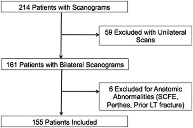
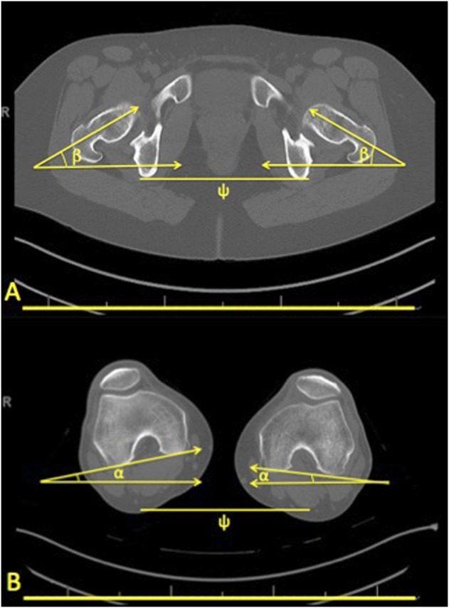
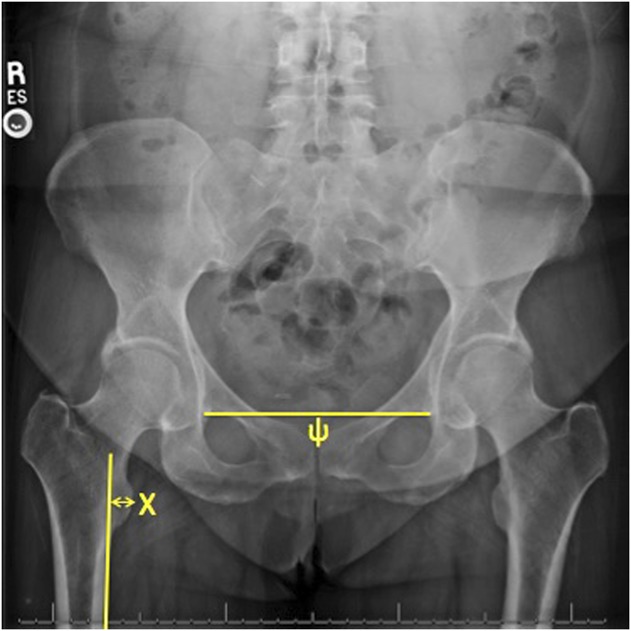
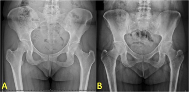
Methods: One hundred fifty-five consecutive patients (72% female and 28% male) with a mean age of 32 years (range, 12-56 years) with a CT scanogram were retrospectively evaluated. Patients were included if CT scanograms had adequate cuts of the proximal and distal femur. Patients were excluded if they had prior hip/femur surgery or anatomic abnormalities of the proximal femur. CT scanogram measurements of femoral rotation were compared with the lesser trochanter profile (distance from the tip of the lesser trochanter to the medial cortex of the femur) measured on weightbearing AP radiographs. These measurements were made by a single fellowship-trained orthopaedic surgeon and repeated for intraobserver reliability testing. Presence of rotational differences based on sex and laterality was assessed and correlation of the difference in lesser trochanter profile to the difference in femoral rotation was determined using a coefficient of determination (r).
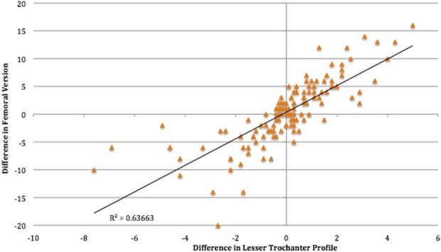
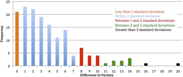
Results: The mean femoral rotation was 10.9° (SD ± 8.8°) of anteversion. Mean right femoral rotation was 11.0° (SD ± 8.9°) and mean left femoral rotation was 10.7° (SD ± 8.7°) with a mean difference of 0.3° (95% confidence interval [CI], -1.7° to 2.3°; p = 0.76). Males had a mean rotation of 9.4°(SD ± 7.7°) and females had a mean rotation of 11.5° (SD ± 9.1°) with a mean difference of 2.1° (95% CI, -0.1° to 4.3°; p = 0.06). Mean lesser trochanter profile was 6.6 mm (SD ± 4.0 mm). Mean right lesser trochanter profile was 6.6 mm (SD ± 3.9 mm) and mean left lesser trochanter profile was 6.5 mm (SD ± 4.0 mm) with a mean difference of 0.1 mm (-0.8 mm to 1.0 mm, p = 0.86). The lesser trochanter profile varied between the sexes; males had a mean of 8.3 mm (SD ± 3.4), and females had a mean of 5.9 mm (SD ± 4.0). The mean difference between sexes was 2.5 mm (1.5-3.4 mm; p < 0.001). The magnitude of the lesser trochanter profile measurement and degree of femoral rotation were positively correlated such that increasing measures of the lesser trochanter profile were associated with increasing amounts of femoral anteversion. The lesser trochanter profile was associated with femoral version in a linear regression model (r = 0.64; p < 0.001). Thus, 64% of the difference in femoral rotation can be explained by the difference in the lesser trochanter profile. Intraobserver reliability for both the femoral version and lesser trochanter profile was noted to be excellent with intraclass correlation coefficients of 0.94 and 0.95, respectively.
Conclusions: This study helps define the normal femoral rotation profile among adults without femoral injury or bone deformity and demonstrated no rotational differences between sexes. The lesser trochanter profile was found to be positively associated with femoral rotation. Increasing and decreasing lesser trochanter profile measurements are associated with increasing and decreasing amounts of femoral rotation, respectively.
Clinical relevance: The lesser trochanter profile can determine the position of the femur in both anteversion and retroversion, supporting its use as a method to restore preinjury femoral rotation after fracture fixation. Although some variability in the rotation between sides may exist, matching the lesser trochanter profile between injured and uninjured femora can help reestablish native rotation.
Conflict of interest statement
All ICMJE Conflict of Interest Forms for authors and
Figures
Comment in
-
CORR Insights®: Is the Lesser Trochanter Profile a Reliable Means of Restoring Anatomic Rotation After Femur Fracture Fixation?Clin Orthop Relat Res. 2018 Jun;476(6):1262-1263. doi: 10.1097/01.blo.0000534690.66772.c8. Clin Orthop Relat Res. 2018. PMID: 29771854 Free PMC article. No abstract available.
References
-
- Bråten M, Terjesen T, Rossvoll I. Femoral anteversion in normal adults. Ultrasound measurements in men and 50 women. Acta Orthop Scand. 1992;63:29–32. - PubMed
-
- Bråten M, Terjesen T, Rossvoll I. Torsional deformity after intramedullary nailing of femoral shaft fractures. Measurement of anteversion angles in 110 patients. J Bone Joint Surg Br. 1993;75:799–803. - PubMed
-
- Bråten M, Terjesen T, Rossvoll I. Femoral shaft fractures treated by intramedullary nailing. A follow-up study focusing on problems related to the method. Injury. 1995;26:379–383. - PubMed
-
- Bråten M, Tveit K, Junk S, Aamodt A, Anda S, Terjesen T. The role of fluoroscopy in avoiding rotational deformity of treated femoral shaft fractures: an anatomical and clinical study. Injury. 2000;31:311–315. - PubMed
-
- Brouwer KJ, Molenaar JC, van Linge B. Rotational deformities after femoral shaft fractures in childhood. A retrospective study 27-32 years after the accident. Acta Orthop Scand. 1981;52:81–89. - PubMed
Publication types
MeSH terms
LinkOut - more resources
Full Text Sources
Other Literature Sources
Medical
