. 2018 Mar;6(2):374-383.
doi: 10.1111/andr.12471. Epub 2018 Feb 22.

High-quality human and rat spermatozoal RNA isolation for functional genomic studies

Affiliations

High-quality human and rat spermatozoal RNA isolation for functional genomic studies

E Bianchi et al. Andrology. 2018 Mar.

Abstract

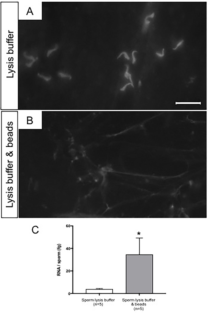
Sperm RNA is a sensitive monitoring endpoint for male reproductive toxicants, and a potential biomarker to assess male infertility and sperm quality. However, isolation of sperm RNA is a challenging procedure due to the heterogeneous population of cells present in the ejaculate, the low yield of RNA per spermatozoon, and the absence of 18S and 28S ribosomal RNA subunits. The unique biology of spermatozoa has created some uncertainty in the field about RNA isolation methods, indicating the need for rigorous quality control checks to ensure reproducibility of data generated from sperm RNA. Therefore, we developed a reliable and effective protocol for RNA isolation from rat and human spermatozoa that delivers highly purified and intact RNA, verified using RNA-specific electrophoretic chips and molecular biology approaches such as RT-PCR and Western blot analysis. The sperm RNA isolation technique was optimized using rat spermatozoa and then adapted to human spermatozoa. Three steps in the sperm isolation procedure, epididymal fluid collection, sperm purification, and spermatozoon RNA extraction, were evaluated and assessed. The sperm RNA extraction methodology consists of collection of rat epididymal fluid with repeated needle punctures of the epididymis, somatic cell elimination using detergent-based somatic cell lysis buffer (SCLB) and the use of RNA isolation Kit. Rat sperm heads are more resistant to disruption than human spermatozoa, necessitating the addition of mechanical lysis with microbeads and heat in the rat protocol, whereas the human sperm protocol only required lysis buffer. In conclusion, this methodology results in reliable and consistent isolation of high-quality sperm RNA. Using this technique will aid in translation of data collected from animal models, and reproducibility of clinical assessment of male factor fertility using RNA molecular biomarkers.

Keywords: RNA; high-quality methodology; human; integrity; rat; spermatozoa.


Figures

Figure 1.
Figure 1.. Rat sperm collection, preparation and RNA isolation.
Procedural diagram for the collection, preparation and isolation of rat sperm RNAs. Rat epididymal fluid samples collected by microdissection (method A) or repeated needle punctures of epididymis (methods B, C, D, E). The whole fluid processed to eliminate somatic cells contamination by somatic cell lysis buffer (methods A, B, C, D) or red blood cell lysis buffer (method E). Sperm cell lysed with only lysis buffer (method C) or lysis buffer, 65°C heat and agitation with 100 μL microbeads (methods D, E). Sperm RNAs isolated using the total RNA isolation kit (methods A, B, C, D, E).
Figure 2.
Figure 2.. Electropherograms of rat sperm RNAs isolated using different collection approaches and preparation procedures.
A) RNA electropherogram profiles of rat sperm collected by the whole epididymal fluid microdissection (method A) and repeated needle punctures of epididymis (method B). B) RNA electropherogram profiles of rat sperm processed with somatic cell lysis buffer (method D) and red blood cell lysis buffer (method E). C) Electropherogram band intensity of 18S and 28S rRNA peaks of rat sperm extracts from whole epididymal fluid collected by microdissection (method A) and repeated needle punctures of epididymis (method B). D) Electropherogram band intensity of 18S and 28S rRNA peaks of rat sperm processed with somatic cell lysis buffer (method D) and red blood cell lysis buffer (method E). 18S and 28S rRNA peaks (red arrows). Data were analyzed using a two-tailed Student’s t-test. *** p-value <0.001, **** p-value <0.0001.
Figure 3.
Figure 3.. Prm2 transcript analysis of rat sperm isolated using different collection and preparation procedures.
A) Prm2 transcript level of whole rat epididymal fluid collected by microdissection (method A) or repeated needle punctures of epididymis (method B). Method A was used as control group. B) Prm2 transcript levels of rat sperm processed with somatic cell lysis buffer (method D) or red blood cell lysis buffer (method E). Method E was used as control group. Data were analyzed by two-tailed Student’s t-test. * p-value <0.05.
Figure 4.
Figure 4.. Comparison of the capability of sperm lysis buffer to dissolve rat sperm heads alone or with microbeads and heat.
A) Microscopic examination of the rat sperm nuclei on lysis buffer (method C) or lysis buffer with microbeads and heat (method D) to assess their ability to completely lyse sperm heads. Bar = 50 μm. B) RNA yield per rat spermatozoon on sperm heads lysed using only lysis buffer or lysis buffer with microbeads and heat. Data were analyzed by two-tailed Student’s t-test. * p-value <0.05.
Figure 5.
Figure 5.. Human sperm collection, preparation and RNA isolation.
A) Procedural diagram for human sperm collection, preparation and RNA isolation. Fresh human semen samples were collected and analyzed according to 2010 WHO guidelines. The ejaculates were immediately washed and somatic cells were lysed with somatic cell lysis buffer (SCLB) to remove non-sperm cells. Sperm RNA was isolated. B) Recovery of RNA per sperm cell after removal of somatic cells via treatment with different dilutions of somatic cell lysis buffer. SLCB A: 0.1% SDS & 0.5% Triton; SLCB B: 0.05% SDS & 0.25% Triton; SLCB C: 0.025% SDS & 0.25% Triton; SLCB D: 0.010% SDS & 0.10% Triton. The RNA yield per sperm was calculated combining large RNA and small RNA of each sperm sample. Sperm purification with SCLB B resulted in a higher RNA yield per sperm compared to the other SCLBs (p=0.015 compared to SCLB A, n.s compared to SCLB C, and p=0.033 compared to SCLB D). Data were analyzed by one-way ANOVA Turkey’s multiple comparisons, p-value < 0.05. C) Recovery of RNA per sperm cell after spermatozoa lysis via sperm lysis buffer or sperm lysis buffer and microbeads treatments. Data were analyzed by two-tailed Student’s t-test.
Figure 6.
Figure 6.. Integrity of human sperm RNA isolated and absence of somatic cells contamination.
A) Bioanalyzer electropherogram of a representative human sperm RNA preparation showing absence of ribosomal RNA and genomic DNA contamination. B) Agarose gel images showing RT-PCR results of five representative human sperm RNA samples (n=5) isolates following the optimized RNA isolation protocol. RNA integrity assessed with PRM2 while somatic cell contamination with CD45 and CDH1. Reference genes UBC and RPLP2 were used.
Figure 7.
Figure 7.. Absence of somatic cell contamination in human sperm samples confirmed by western blot analysis.
Western blot analysis was performed to assess CDH1 and CD45 protein levels in human sperm pellet. 30 εg of cell lysates proteins were loaded onto a 7% and 12% acrylamide gels. A) MCF7 cell lysate admixed to the human sperm lysate. Lysate of 7.50 × 10⁴ MCF7 cells (MCF7, 30 εg of MCF7 lysate); lysates of 3.70 × 10⁴ MCF7 cells and 1.17 ×10⁷ sperm cells (1 MCF7:1 Sperm, 15 εg of MCF7 lysate and 15 εg of sperm lysate); lysates of 7.50 × 10³ MCF7 cells and 1.80 ×10⁷ sperm cells (1 MCF7:10 Sperm, 3 εg of MCF7 lysate and 27 εg of sperm lysate); lysates of 2.30 ×10⁷ sperm cells (Sperm, 30 εg of sperm lysate). B) Jurkat cell lysate admixed to the human sperm lysate. Lysate of 4.90 × 10⁴ Jurkat cells (Jurkat, 30 εg of Jurkat lysate); lysates of 2.44 × 10⁴ Jurkat cells and 5.70 ×10⁶ sperm cells (1 Jurkat:1 Sperm, 15 εg of Jurkat lysate and 15 εg of sperm lysate); lysates of 4.88 × 10³ Jurkat cells and 1.02 ×10⁷ sperm cells (1 Jurkat:10 Sperm, 3 εg of Jurkat lysate and 27 εg of sperm lysate); lysates of 1.13 ×10⁷ sperm cells (Sperm, 30 εg of sperm lysate).

References

    1. Inhorn MC, Patrizio P. Infertility around the globe: new thinking on gender, reproductive technologies and global movements in the 21st century. Hum Reprod Update 2015; 21: 411–26. - PubMed
    1. Jarow JP, Sharlip ID, Belker AM, Lipshultz LI, Sigman M, Thomas AJ, et al. Best practice policies for male infertility. J Urol 2002; 167: 2138–44. - PubMed
    1. Gottardo F, Kliesch S, World Health O. [Semen analysis: spermiogram according to WHO 2010 criteria]. Urologe A 2011; 50: 101–8. - PubMed
    1. Lipshultz LI, Lamb DJ. Risk of transmission of genetic diseases by assisted reproduction. Nat Clin Pract Urol 2007; 4: 460–1. - PMC - PubMed
    1. Kierszenbaum AL. Mammalian spermatogenesis in vivo and in vitro: a partnership of spermatogenic and somatic cell lineages. Endocr Rev 1994; 15: 116–34. - PubMed

Publication types

LinkOut - more resources