. 2018 Mar 16;46(5):2560-2572.
doi: 10.1093/nar/gky107.

The H-subunit of the restriction endonuclease CglI contains a prototype DEAD-Z1 helicase-like motor

Affiliations

The H-subunit of the restriction endonuclease CglI contains a prototype DEAD-Z1 helicase-like motor

Paulius Toliusis et al. Nucleic Acids Res. .

Abstract

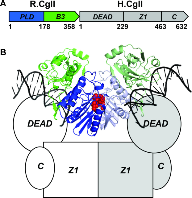
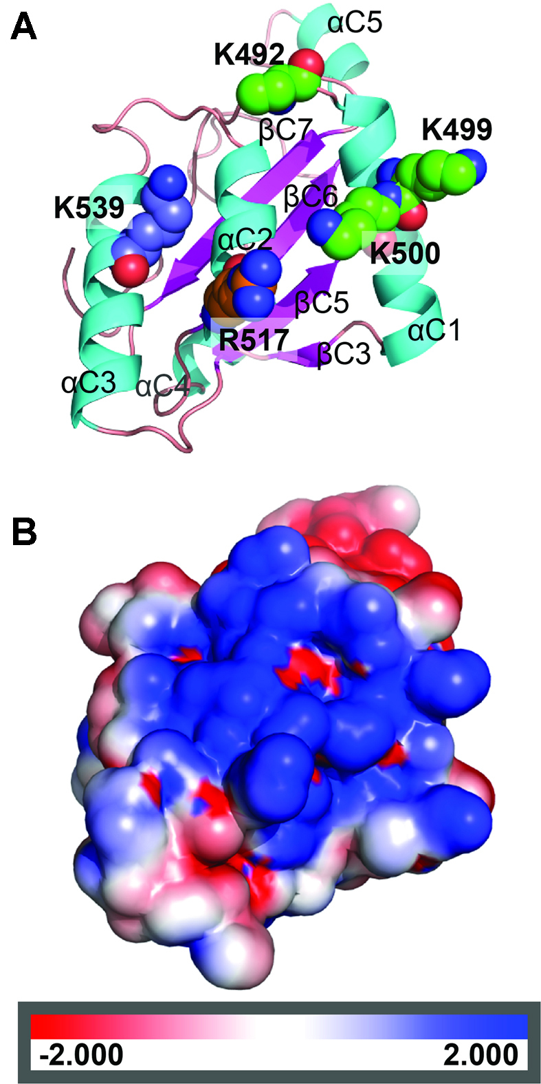
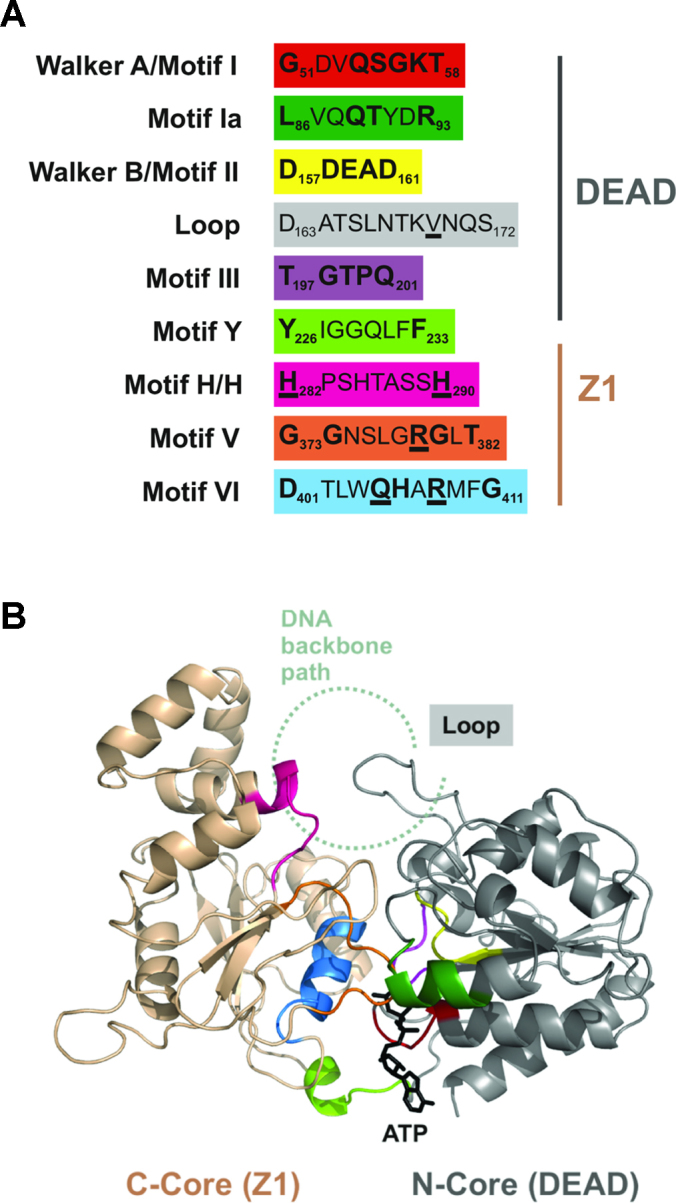
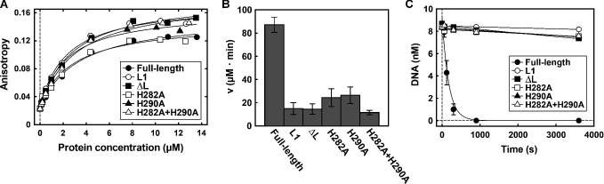
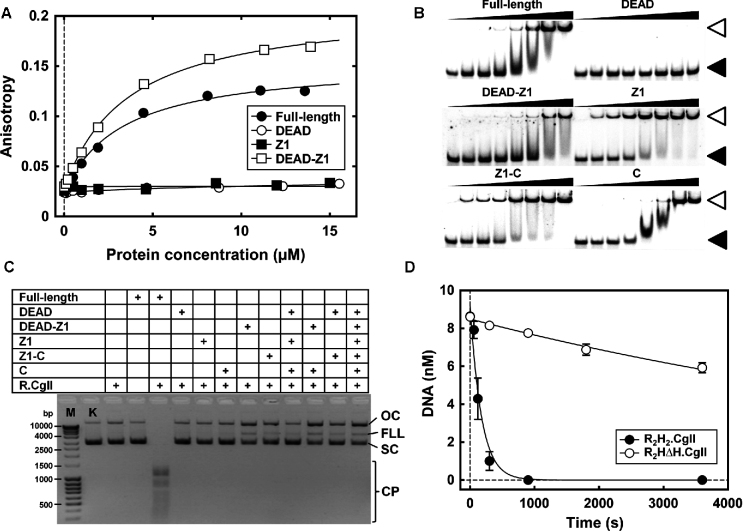
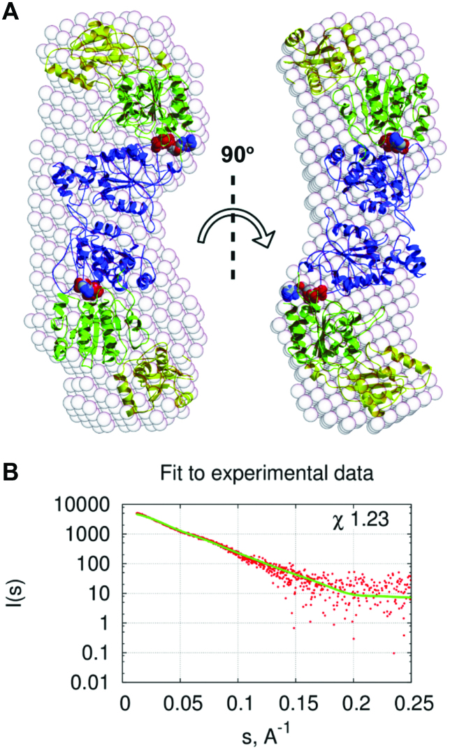
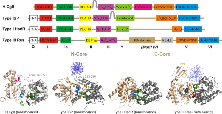
CglI is a restriction endonuclease from Corynebacterium glutamicum that forms a complex between: two R-subunits that have site specific-recognition and nuclease domains; and two H-subunits, with Superfamily 2 helicase-like DEAD domains, and uncharacterized Z1 and C-terminal domains. ATP hydrolysis by the H-subunits catalyses dsDNA translocation that is necessary for long-range movement along DNA that activates nuclease activity. Here, we provide biochemical and molecular modelling evidence that shows that Z1 has a fold distantly-related to RecA, and that the DEAD-Z1 domains together form an ATP binding interface and are the prototype of a previously undescribed monomeric helicase-like motor. The DEAD-Z1 motor has unusual Walker A and Motif VI sequences those nonetheless have their expected functions. Additionally, it contains DEAD-Z1-specific features: an H/H motif and a loop (aa 163-aa 172), that both play a role in the coupling of ATP hydrolysis to DNA cleavage. We also solved the crystal structure of the C-terminal domain which has a unique fold, and demonstrate that the Z1-C domains are the principal DNA binding interface of the H-subunit. Finally, we use small angle X-ray scattering to provide a model for how the H-subunit domains are arranged in a dimeric complex.


Figures

Figure 1.
Figure 1.
The restriction endonuclease CglI from Corynebacterium glutamicum. (A) Domain organisation of R- and H.CglI proteins. R.CglI (NCgl1704) has PLD-superfamily nuclease and B3-like DNA binding domains. H.CglI (NCgl1705) has a predicted SF2 helicase/ATPase domain (DEAD) linked to uncharacterized Z1-superfamily and C-terminal domains. Numbers represent predicted amino acid positions of domain boundaries. (B) A possible model of the R2H2.CglI complex (6,79). The R.CglI dimer is presented by the structure of the homologous R.NgoAVII (PDB ID 4RCT) bound to the cognate DNA via the B3 domains (PDB ID 4RD5). Two R.NgoAVII PLD domains from separate subunits (coloured blue and light blue) dimerise to form single active site (nuclease active site residues H104 and K106 are coloured red). B3-like domains (coloured green and light green) are found in plant transcription factors and recognise the 5′-GCCGC-3′ target. DNA is shown as black ribbon. H.CglI contains the DEAD, Z1 and C-terminal domains (separate subunits are white or light grey).
Figure 2.
Figure 2.
Crystal structure of the H.CglI C-terminal domain. (A) Ribbon representation of the C-terminal domain with possible aa responsible for DNA binding. The positively charged residues subjected to mutagenesis are shown as spacefill. (B) Electrostatic surface potential of the C-terminal domain (the protein orientation is as in (A)).
Figure 3.
Figure 3.
Putative helicase motifs and model for the DEAD-Z1 helicase of H.CglI. (A) H.CglI helicase motifs identified from the alignment of ORFs related to the H-subunit (Supplementary Figure S5). The loop was identified from the model structure shown in panel B. Residues in bold are part of the DEAD-Z1 consensus sequences. Underlining indicates that a mutation was made at that amino acid in this study. (B) Model of the DEAD-Z1 helicase of H.CglI (aa 22 – aa 447). The DEAD domain is coloured in grey, the Z1 domain in gold. The motifs are coloured as in panel A. ATP is shown as a stick model. An approximate location of the path of a DNA backbone was constructed by similarity to the SsoRad54-DNA complex (49), PDB ID 1Z63.
Figure 4.
Figure 4.
Enzyme properties of Loop and H/H motif mutants. (A) Mant-ATP binding was studied by fluorescence anisotropy measurements. Data points are fitted to a hyperbolic function. (B) Rates of ATP hydrolysis. Reactions contained 0.62 nM phage λ DNA (contains 181 CglI sites), 10 nM H.CglI, 200 nM R.CglI, 2 mM ATP. (C) Rate of cleavage of the first strand (nicking) of one-site supercoiled DNA. Data points are fitted to a single exponential decay function. The fitted values for panels A-C are presented in Supplementary Tables S4–S6. All data is the average of three or more reactions, with the error bars ± SD where shown.
Figure 5.
Figure 5.
Functional analysis of the domains of H.CglI. (A) Mant-ATP binding by the full-length H.CglI and its isolated domains was studied by fluorescence anisotropy measurements. Data points were fitted to a hyperbolic function. (B) DNA binding by the full-length H.CglI and its domains measured using an EMSA. The reactions contained 1 nM of the 33P-labelled oligoduplex and the proteins indicated at increasing concentrations (nM): 0, 50, 100, 200, 500, 1000, 2000 and 5000 (shown as wide black triangles). Images of separate native polyacrylamide gels are shown, obtained by Fujifilm FLA-5100 fluorescent image analyzer and the OptiQuant 3.0 software. Filled and open triangles correspond to the mobility of the free DNA and a protein–DNA complex, respectively. (C) Cleavage of the 21-site pBR322 plasmid DNA by the mixtures of the R.CglI and H.CglI or its domains, as indicated, for 1 h at 37°C. M – marker (DNA fragments of known length in bp), K – control (pBR322 plasmid DNA), SC – supercoiled circular DNA, OC – open circular DNA (‘nicked’), FLL – full-length linear DNA, CP – cleavage products at multiple sites. (D) Rate of cleavage of the first strand (nicking) of one-site supercoiled DNA by the wt R2H2.CglI and R2HΔH.CglI (lacking a single C-terminal domain within H.CglI) complexes. Data points are fitted to a single exponential function. The fitted values for panels A, B and C are presented in Supplementary Tables S3, S4 and S6. All data points are the average of 3 or more reactions, with the error bars ± SD.
Figure 6.
Figure 6.
The H.CglI dimer modeling using SAXS data. (A) Domain movement (SASREF) model compared with dummy atom model. Dummy atom model of the full-length H.CglI calculated by DAMMIN is shown as transparent spheres. The DEAD and Z1-domains were taken from the DEAD-Z1 model and are coloured green and blue, respectively. The C-terminal domains from the crystal structure (PDB ID 6F1S) are coloured yellow; ATP is shown in red CPK. (B) Fit of the SASREF model (green line) to SAXS data (red points).
Figure 7.
Figure 7.
Comparison of the helicase-like domains of Types I, ISP and III restriction enzymes. Helicase consensus motifs are shown based on the work here and (61,78,80,81). The Type IIIA consensus sequences are presented. Loop regions and the Type III PIN domain are indicated. ‘h’ represents a hydrophobic residue, ‘x’ represents <85% consensus, ‘…’ represents a variable amino acid spacing. The N- and C-core helicase-like domain structures are shown for (from left to right): H.CglI, Type ISP LlaBIII (PDB ID 4XQK, (78)), Type I HsdR subunit from EcoR124I (PDB ID 2W00, (82)), and Type III Res subunit from EcoP15I (PDB ID 4ZCF, (81)). Helicase motifs are coloured as in the upper panel. The expected position of Motif IV is indicated, although it is less easily identified in restriction enzymes (61). CglI, Type ISP and Type I enzymes are dsDNA translocases that consumes ∼1 ATP/bp (5,77,83). Type III enzymes are molecular switches that consumes 10–20 bp, change conformation, and then move on DNA by thermally-driven DNA sliding (3).

Similar articles

Cited by

References

    1. Wu C.G., Spies M.. Overview: what are helicases. Adv. Exp. Med. Biol. 2013; 973:1–16. - PubMed
    1. Clapier C.R., Iwasa J., Cairns B.R., Peterson C.L.. Mechanisms of action and regulation of ATP-dependent chromatin-remodelling complexes. Nat. Rev. Mol. Cell Biol. 2017; 18:407–422. - PMC - PubMed
    1. Schwarz F.W., Toth J., van Aelst K., Cui G., Clausing S., Szczelkun M.D., Seidel R.. The helicase-like domains of type III restriction enzymes trigger long-range diffusion along DNA. Science. 2013; 340:353–356. - PMC - PubMed
    1. Szczelkun M.D. Roles for helicases as ATP-dependent molecular switches. Adv. Exp. Med. Biol. 2013; 767:225–244. - PubMed
    1. Toliusis P., Zaremba M., Silanskas A., Szczelkun M.D., Siksnys V.. CgII cleaves DNA using a mechanism distinct from other ATP-dependent restriction endonucleases. Nucleic Acids Res. 2017; 45:8435–8447. - PMC - PubMed

Publication types

MeSH terms

LinkOut - more resources