. 2018 Feb 23;17(1):65.
doi: 10.1186/s12943-018-0809-x.

Transcriptional profiles of different states of cancer stem cells in triple-negative breast cancer

Affiliations

Transcriptional profiles of different states of cancer stem cells in triple-negative breast cancer

Mingshan Liu et al. Mol Cancer. .

Abstract

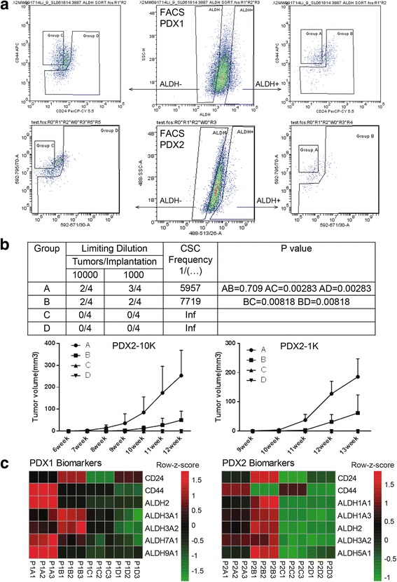
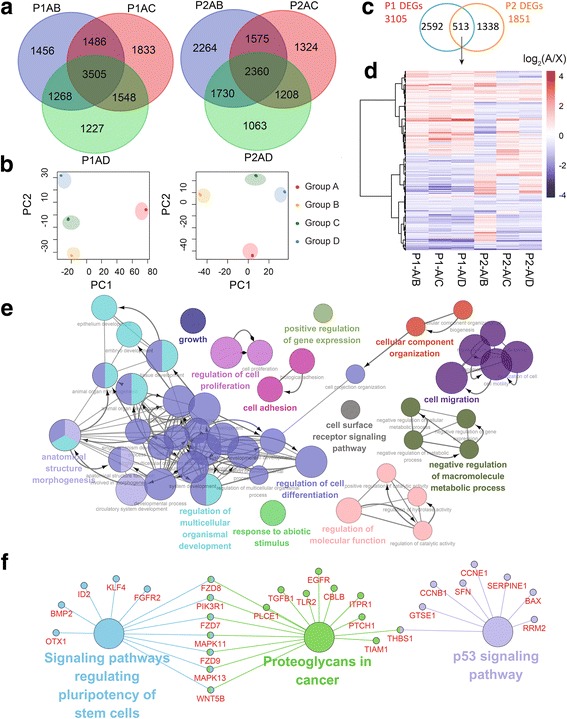
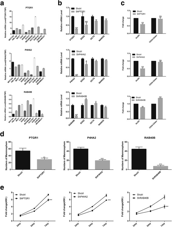
Breast cancer stem cells (BCSCs) are thought to be responsible for tumor initiation, metastasis and relapse. Our group and others have described markers useful in isolating BCSCs just as aldehyde dehydrogenase positive (ALDH+) or CD24-CD44+. In fact, cells which simultaneously express both sets of markers have the highest tumor initiating capacity. Although the transcriptomic profile of cells expressing each BCSC marker alone has been reported, the profile of the most tumorigenic population expressing both sets of markers has not. Here we used the biomarker combination of ALDH and CD24/CD44 to sort four populations isolated from triple-negative breast cancer (TNBC) patient-derived xenografts, and performed whole-transcriptome sequencing on each population. We systematically compared the profiles of the three states of BCSCs (ALDH+CD24-CD44+, ALDH+non-CD24-CD44+ and ALDH-CD24-CD44+) to that of the differentiated tumor cells (ALDH-non-CD24-CD44+). For the first time, we compared the ALDH+CD24-CD44+ BCSCs with the other two BCSC populations. In ALDH+CD24-CD44+ BCSCs, we identified P4HA2, PTGR1 and RAB40B as potential prognostic markers, which were virtually related to the status of BCSCs and tumor growth in TNBC cells.

Keywords: Cancer stem cells; Triple-negative breast cancer; Whole-transcriptome sequencing.


Conflict of interest statement

Ethics approval and consent to participate

All mouse experiments were conducted in accordance with standard operating procedures approved by the University Committee on the Use and Care of Animals at University of Science and Technology of China.

Consent for publication

Not applicable.

Competing interests

The authors declare that they have no competing interests.

Publisher’s Note

Springer Nature remains neutral with regard to jurisdictional claims in published maps and institutional affiliations.

Figures

Fig. 1
Fig. 1
Isolation and characterization of the four cell populations from PDXs. (a) The flow charts of ALDH, CD24 and CD44 for PDX1 and PDX2 by fluorescent activated cell sorting. We isolated four groups based on biomarker combinations of ALDH and CD24/CD44. (b) The limiting dilutions of cells obtained from PDX2 (VARI068) were implanted in the fourth fat pads of NOD-SCID mice. The tumor growth for each group was monitored and calculated weekly, and the CSC frequency for the group A, B, C, D was calculated based on the website http://bioinf.wehi.edu.au/software/elda/. (c) The expression of BCSC biomarkers ALDH, CD24 and CD44 in each sorted group. We compared each group with the following criteria: 1) CD24: A < B, C < D; 2) CD44: A > B, C > D; 3) ALDH: A/B > C/D. PDXs have different expressions of ALDH isoforms. P1: PDX1; P2: PDX2
Fig. 2
Fig. 2
The unique DEGs of ALDH+CD24CD44+ BCSCs. (a) The Venn diagrams of the DEGs between ALDH+CD24CD44+ BCSCs (group A) and other three groups with fold change set 1.2. (b) The principal component analysis (PCA) plots of DEGs in two PDXs. (c) The intersection set of DEGs after filtered by PCA in two PDXs. (d) The intersected 513 DEGs in two PDXs. (e) The GO analysis based on biological processes of the 513 DEGs visualized by Apps ClueGO v2.3.2 of Cytoscape v3.4.0 with network specificity set Global. (f) The KEGG pathway analysis of the 513 DEGs visualized by Cytoscape v3.4.0 with network specificity set medium
Fig. 3
Fig. 3
Functional analysis of potential prognostic genes. (a) The expressions of PTGR1, P4HA2 and RAB40B was variable across different breast cell lines, including: 1) normal mammary gland cell lines, MCF10A and HBL100; 2) luminal breast cancer cell lines, MCF7 and T47D (ER+PRHER2); 3) HER2+ breast cancer cell lines (ERPRHER2+) containing SKBR3, BT474; 4) Basal-like/TNBC (ERPRHER2) breast cancer cell lines, such as MDA-MB-468, HCC1937, SUM149, SUM159 and MDA-MB-231. (b) The expressions of CSC-related genes (ShPTGR1, ShP4HA2 and ShRAB40B) in the knockdown and the control (Shctrl) TNBC cell line SUM149. (c) The fold change for the proportion of each BCSC state in knockdown cells vs. Shctrl cells as assessed by fluorescent activated cell sorting. (d) The mammosphere formed in Shctrl cells and knockdown cells accessed by mammosphere formation assay. (e) The fold change for cell proliferation of knockdown SUM149 cells vs. Shctrl SUM149 cells as assessed by MTT assay . *, P < 0.05; **, P < 0.01; ***, P < 0.001; ns, not significant (compared with the corresponding Shctrl group). Error bars, mean ± SD

References

    1. Bianchini G, Balko JM, Mayer IA, Sanders ME, Gianni L. Triple-negative breast cancer: challenges and opportunities of a heterogeneous disease. Nat Rev Clin Oncol. 2016;13:674–690. doi: 10.1038/nrclinonc.2016.66. - DOI - PMC - PubMed
    1. Malorni L, Shetty PB, De Angelis C, Hilsenbeck S, Rimawi MF, Elledge R, Osborne CK, De Placido S, Arpino G. Clinical and biologic features of triple-negative breast cancers in a large cohort of patients with long-term follow-up. Breast Cancer Res Treat. 2012;136:795–804. doi: 10.1007/s10549-012-2315-y. - DOI - PMC - PubMed
    1. Beck B, Blanpain C. Unravelling cancer stem cell potential. Nat Rev Cancer. 2013;13:727–738. doi: 10.1038/nrc3597. - DOI - PubMed
    1. Nguyen LV, Vanner R, Dirks P, Eaves CJ. Cancer stem cells: an evolving concept. Nat Rev Cancer. 2012;12:133–143. doi: 10.1038/nrc3184. - DOI - PubMed
    1. Lawson DA, Bhakta NR, Kessenbrock K, Prummel KD, Yu Y, Takai K, et al. Single-cell analysis reveals a stem-cell program in human metastatic breast cancer cells. Nature. 2015;526:131–135. doi: 10.1038/nature15260. - DOI - PMC - PubMed

Publication types

Substances

LinkOut - more resources