. 2018 Feb 23;11(1):26.
doi: 10.1186/s13045-018-0577-5.

Oncoprotein HBXIP enhances HOXB13 acetylation and co-activates HOXB13 to confer tamoxifen resistance in breast cancer

Affiliations

Oncoprotein HBXIP enhances HOXB13 acetylation and co-activates HOXB13 to confer tamoxifen resistance in breast cancer

Bowen Liu et al. J Hematol Oncol. .

Abstract

Background: Resistance to tamoxifen (TAM) frequently occurs in the treatment of estrogen receptor positive (ER+) breast cancer. Accumulating evidences indicate that transcription factor HOXB13 is of great significance in TAM resistance. However, the regulation of HOXB13 in TAM-resistant breast cancer remains largely unexplored. Here, we were interested in the potential effect of HBXIP, an oncoprotein involved in the acceleration of cancer progression, on the modulation of HOXB13 in TAM resistance of breast cancer.

Methods: The Kaplan-Meier plotter cancer database and GEO dataset were used to analyze the association between HBXIP expression and relapse-free survival. The correlation of HBXIP and HOXB13 in ER+ breast cancer was assessed by human tissue microarray. Immunoblotting analysis, qRT-PCR assay, immunofluorescence staining, Co-IP assay, ChIP assay, luciferase reporter gene assay, cell viability assay, and colony formation assay were performed to explore the possible molecular mechanism by which HBXIP modulates HOXB13. Cell viability assay, xenograft assay, and immunohistochemistry staining analysis were utilized to evaluate the effect of the HBXIP/HOXB13 axis on the facilitation of TAM resistance in vitro and in vivo.

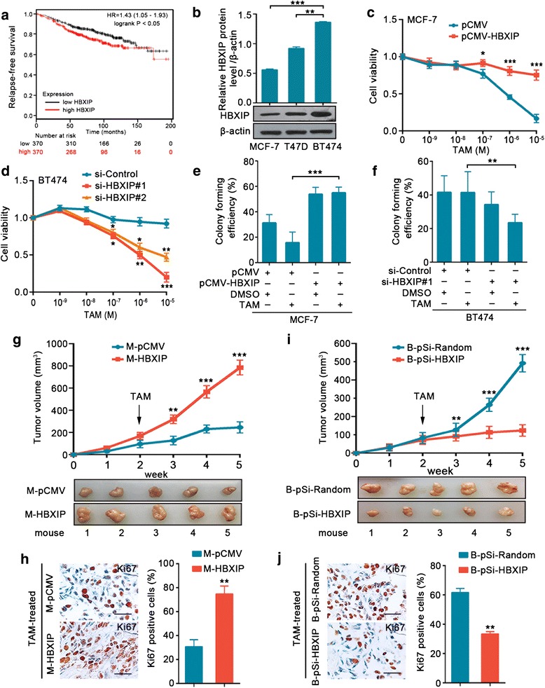
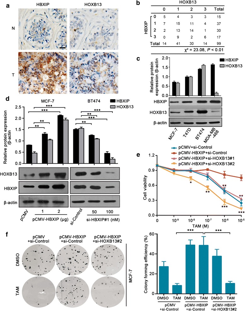
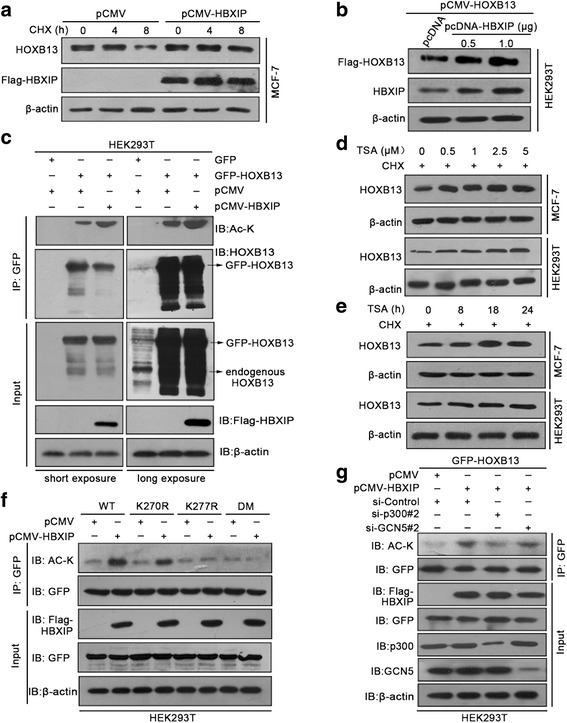
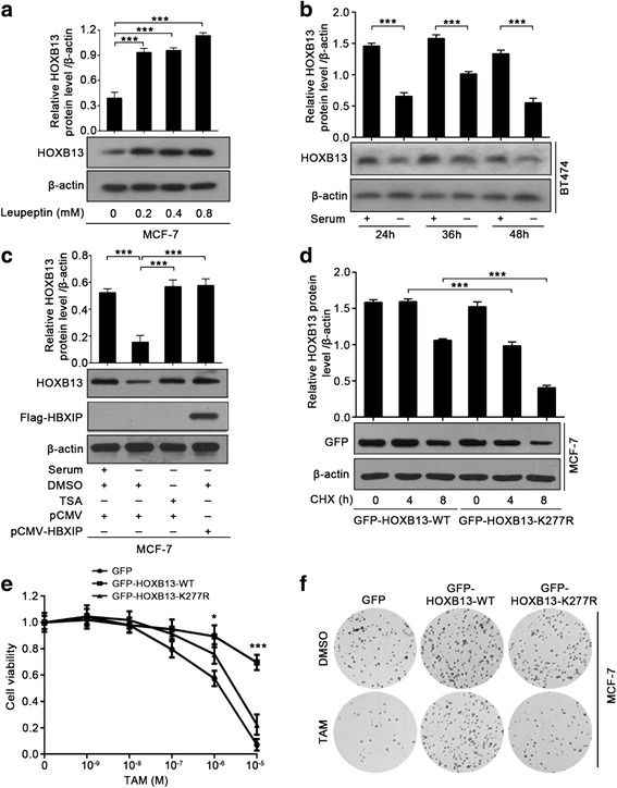
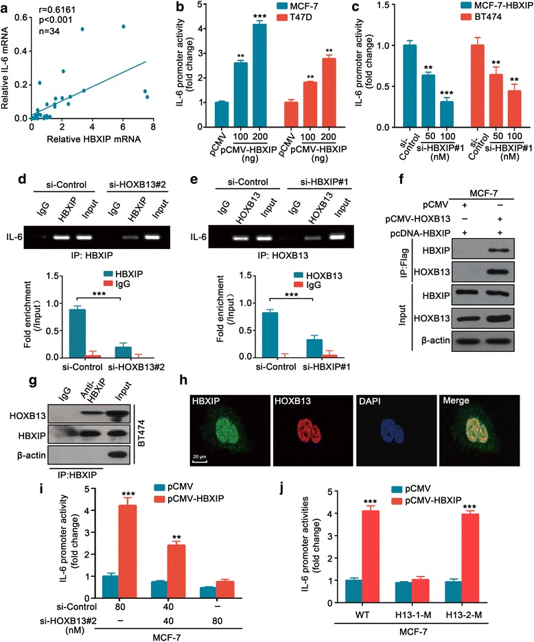
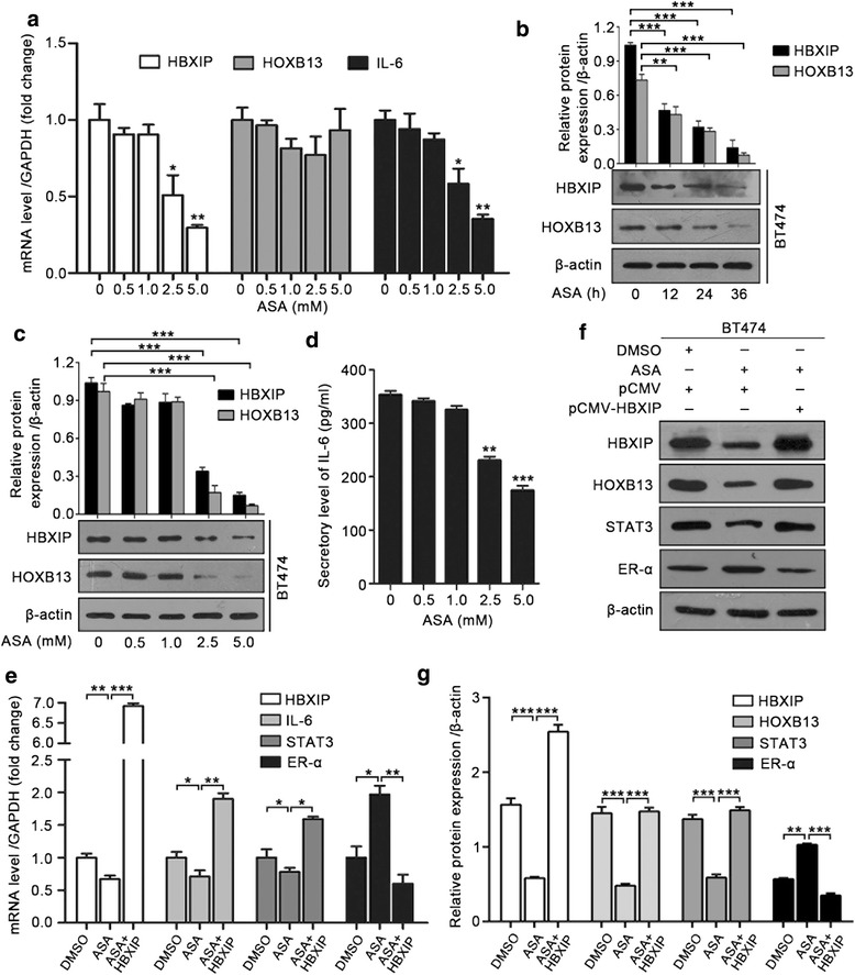
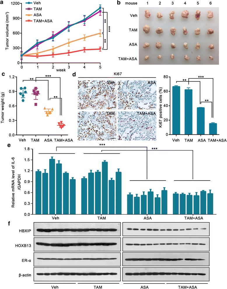
Results: The analysis of the Kaplan-Meier plotter and the GEO dataset showed that mono-TAM-treated breast cancer patients with higher HBXIP expression levels had shorter relapse-free survivals than patients with lower HBXIP expression levels. Overexpression of HBXIP induced TAM resistance in ER+ breast cancer cells. The tissue microarray analysis revealed a positive association between the expression levels of HBXIP and HOXB13 in ER+ breast cancer patients. HBXIP elevated HOXB13 protein level in breast cancer cells. Mechanistically, HBXIP prevented chaperone-mediated autophagy (CMA)-dependent degradation of HOXB13 via enhancement of HOXB13 acetylation at the lysine 277 residue, causing the accumulation of HOXB13. Moreover, HBXIP was able to act as a co-activator of HOXB13 to stimulate interleukin (IL)-6 transcription in the promotion of TAM resistance. Interestingly, aspirin (ASA) suppressed the HBXIP/HOXB13 axis by decreasing HBXIP expression, overcoming TAM resistance in vitro and in vivo.

Conclusions: Our study highlights that HBXIP enhances HOXB13 acetylation to prevent HOXB13 degradation and co-activates HOXB13 in the promotion of TAM resistance of breast cancer. Therapeutically, ASA can serve as a potential candidate for reversing TAM resistance by inhibiting HBXIP expression.

Keywords: Acetylation; Breast cancer; Chaperone-mediated autophagy; HBXIP; HOXB13; Tamoxifen resistance.


Conflict of interest statement

Ethics approval and consent to participate

This study was approved by the Research Ethics Committee of Nankai University, and written informed consent was obtained from all of the patients.

Consent for publication

Not applicable.

Competing interests

The authors declare that they have no competing interests.

Publisher’s Note

Springer Nature remains neutral with regard to jurisdictional claims in published maps and institutional affiliations.

Figures

Fig. 1
Fig. 1
HBXIP contributes to TAM resistance in breast cancer. a Relapse-free survival analysis of TAM-treated ER+ breast cancer patients by the Kaplan-Meier plotter online resource (http://kmplot.com/analysis/). The plot was generated according to the HBXIP expression level (log-rank P = 0.021). b Immunoblotting analysis of HBXIP in MCF-7, T47D, and BT474 cells (lower panel) and the quantification of the intensity relative to β-actin (upper panel). c, d Cell viability assay in MCF-7 (c) and BT474 (d) cells treated with corresponding doses of TAM after being transiently transfected with the indicated plasmids or siRNAs. e, f Colony forming efficiencies of MCF-7 (e) and BT474 (f) cells treated with DMSO or TAM (1 μM) after being transiently transfected with the indicated plasmids or siRNA. g, h Growth curve and imaging (g), Ki67 (a cell proliferation marker) staining by IHC assay and the statistics of Ki67 positive cells (h) of the xenograft tumors derived from MCF-7-pCMV (called M-pCMV) or MCF-7-HBXIP (called M-HBXIP) cells (each group, n = 5). Scale bar, 50 μm. i, j Growth curve and imaging (i), Ki67 staining by IHC assay and the statistics of Ki67 positive cells (j) of the xenograft tumors derived from BT474-pSilencer-Random (called B-pSi-Random) or BT474-pSilencer-HBXIP (called B-pSi-HBXIP) cells (each group, n = 5). Scale bar, 50 μm. All experiments were repeated at least three times. Error bars represent ± SD. *P < 0.05, **P < 0.01, and ***P < 0.001 by two-tailed Student’s t test
Fig. 2
Fig. 2
HBXIP induces TAM resistance by increasing the protein level of HOXB13. a IHC staining of HBXIP and HOXB13 in normal breast tissues (N) and breast carcinomas (T) from ER+ breast tissue microarray. Scale bar, 20 μm. b The association between HBXIP and HOXB13 expression levels in the abovementioned tissue microarray was statistically analyzed by Pearson chi-square independence test, χ2 = 23.08, P < 0.01. c Immunoblotting analysis of HBXIP and HOXB13 in different breast cancer cell lines (lower panel). The upper panel is the quantification of the intensity relative to β-actin. MDA-MB-468 is a triple-negative breast cancer cell line. d Immunoblotting analysis of HOXB13 in MCF-7 and BT474 cells transiently transfected with the indicated plasmids or siRNA (lower panel). The upper panel is the quantification of the intensity relative to β-actin. e Cell viability assay in MCF-7 cells treated with indicated concentrations of TAM after being transiently transfected with the displayed plasmids or siRNAs. Error bars represent ± SD. *P < 0.05, **P < 0.01, and ***P < 0.001 (HBXIP compared with HBXIP+si-HOXB13) by two-tailed Student’s t test. f A colony photograph and the colony forming efficiency of MCF-7 cells treated with DMSO or TAM (1 μM) after being transiently transfected with the displayed plasmids or siRNA. All experiments were repeated at least three times. Error bars represent ± SD. *P < 0.05, **P < 0.01, and ***P < 0.001 by two-tailed Student’s t test
Fig. 3
Fig. 3
HBXIP enhances acetylation of HOXB13 at K277 site via acetylase p300. a Immunoblotting analysis of HOXB13 in MCF-7 cells time-dependently treated with 100 μg/ml cycloheximide (CHX) after being transiently transfected with the indicated plasmids. b Immunoblotting analysis of exogenous Flag-HOXB13 in HEK293T cells. The cells were transiently transfected with pCMV-HOXB13 accompanied by pcDNA or pcDNA-HBXIP. The protein level of Flag-HOXB13 was examined by the anti-Flag antibody. c Acetylation level of HOXB13 was detected by a Co-IP assay using GFP-beads and an immunoblotting analysis performed with the anti-acetyl-lysine antibody in HEK293T cells. The cells were transiently transfected with the indicated plasmids. The protein levels of HBXIP and HOXB13 were examined by anti-Flag or anti-HOXB13 antibodies, respectively. The left and right panels are identical results that differ in exposure time. d, e Immunoblotting analysis of HOXB13 in MCF-7 cells and HEK293T cells treated with 100 μg/ml CHX along with different concentrations of trichostatin A (TSA) for 18 h (d) or 1 μM TSA (e) for the indicated time points. f Acetylation level of HOXB13 was detected by a Co-IP assay using GFP-beads and an immunoblotting analysis performed with the anti-acetyl-lysine antibody in HEK293T cells. The cells were separately transiently transfected with GFP-HOXB13-WT, GFP-HOXB13-K270R, GFP-HOXB13-K277R, or GFP-HOXB13-DM along with pCMV or pCMV-HBXIP. The protein levels of HBXIP and HOXB13 were examined by anti-Flag or anti-GFP antibodies, respectively. g Acetylation level of HOXB13 was detected by a Co-IP assay using GFP-beads and an immunoblotting analysis with the anti-acetyl-lysine antibody in HEK293T cells. The cells were transiently transfected with GFP-HOXB13-WT along with the indicated plasmids or siRNAs. The protein levels of HBXIP and HOXB13 were separately examined by anti-Flag or anti-GFP antibodies. All experiments were repeated at least three times
Fig. 4
Fig. 4
HBXIP-enhanced acetylation of HOXB13 stabilizes HOXB13 in the facilitation of TAM resistance. a Immunoblotting analysis of HOXB13 in MCF-7 cells treated with the indicated concentrations of leupeptin for 36 h (lower panel). The upper panel is the quantification of the intensity relative to β-actin. b Immunoblotting analysis of HOXB13 in BT474 cells cultured with serum-supplemented or serum-free media for the indicated time courses (lower panel). The upper panel is the quantification of the intensity relative to β-actin. c Immunoblotting analysis of HOXB13 in MCF-7 cells cultured with serum-supplemented or serum-free media for 48 h along with DMSO or TSA (1 μM) (lower panel). Before that, the cells were transiently transfected with pCMV or pCMV-HBXIP (1.5 μg). The protein level of HBXIP was determined by the anti-Flag antibody. The upper panel is the quantification of the intensity relative to β-actin. d Immunoblotting analysis of GFP-HOXB13 in MCF-7 cells time-dependently treated with 100 μg/ml CHX after being transiently transfected with GFP-HOXB13-WT or GFP-HOXB13-K277R (lower panel). The protein level of GFP-HOXB13 was determined by the anti-GFP antibody. The upper panel is the quantification of the intensity relative to β-actin. e Cell viability assay with MCF-7 cells treated with the indicated concentrations of TAM after being transiently transfected with the displayed plasmids. Error bars represent ± SD. *P < 0.05 and ***P < 0.001 (GFP-HOXB13-WT compared with GFP-HOXB13-K277R) by two-tailed Student’s t test. f A colony photograph of MCF-7 cells treated with DMSO or TAM (1 μM) after being transiently transfected with the displayed plasmids. All experiments were repeated at least three times. Error bars represent ± SD. *P < 0.05 and ***P < 0.001 by two-tailed Student’s t test
Fig. 5
Fig. 5
HBXIP co-activates HOXB13 to stimulate IL-6 transcription. a Relative mRNA levels of HBXIP and IL-6 in 34 ER+ clinical breast tumor tissues examined by qRT-PCR assay. The correlation between HBXIP and IL-6 mRNA levels was determined by Pearson’s correlation coefficient. b, c The luciferase reporter gene assay of IL-6 promoter activity in MCF-7 and T47D cells (b) and MCF-7-HBXIP and BT474 cells (c). The cells were transiently transfected with the indicated plasmids or siRNA. The luciferase activities were measured after transfection for 24 h. d ChIP assay in BT474 cells immunoprecipitated with anti-HBXIP antibody or control IgG after being transiently transfected with the si-control or si-HOXB13#2. The lower panel shows the quantitative enrichment data of the IL-6 promoter analyzed by qPCR and normalized against the input. e A similar assay as in d but immunoprecipitated with the anti-HOXB13 antibody or control IgG after being transiently transfected with the si-control or si-HBXIP#1. f Interaction of Flag-HOXB13 with HBXIP was analyzed by Co-IP assay in MCF-7 cells. The cells were transiently transfected with pCMV or pCMV-HOXB13 along with pcDNA-HBXIP. g Interaction of endogenous HBXIP with HOXB13 was examined by Co-IP assay in BT474 cells. h Co-localization of endogenous HBXIP and HOXB13 in BT474 cells was examined by confocal microscopy. Scale bar, 20 μm. i Luciferase reporter gene assay of IL-6 promoter activity in MCF-7 cells transiently transfected with pCMV or pCMV-HBXIP along with the indicated si-control or si-HOXB13#2. j Luciferase reporter gene assay of IL-6 promoter activities in MCF-7 cells. The cells were transiently transfected with pCMV or pCMV-HBXIP along with the IL-6 promoter (WT) or constructs with mutated binding sites of HOXB13-site1 (− 270/− 251, called H13-1-M) or HOXB13-site2 (− 135/− 121, called H13-2-M). All experiments were repeated at least three times. Error bars represent ± SD. **P < 0.01 and ***P < 0.001 by two-tailed Student’s t test
Fig. 6
Fig. 6
ASA suppresses HBXIP/HOXB13 axis by reducing HBXIP expression. a qRT-PCR assay of HBXIP, HOXB13, and IL-6 in BT474 cells treated with the displayed doses of ASA for 24 h. b Immunoblotting analysis of HBXIP and HOXB13 in BT474 cells treated with 2.5 mM ASA for the indicated time points (lower panel). The upper panel is the quantification of the intensity relative to β-actin. c Immunoblotting analysis of HBXIP and HOXB13 in BT474 cells treated with different concentrations of ASA for 24 h (lower panel). The upper panel is the quantification of the intensity relative to β-actin. d ELISA of IL-6 secretion in MCF-7-HBXIP cells treated with the indicated doses of ASA for 24 h. e qRT-PCR assay of HBXIP, IL-6, STAT3, and ER-α in BT474 cells treated with DMSO or ASA (2.5 mM) for 24 h after being transiently transfected with pCMV or pCMV-HBXIP. f Immunoblotting analysis of HBXIP, HOXB13, STAT3, and ER-α in BT474 cells treated with DMSO or ASA (2.5 mM) for 24 h after being transiently transfected with pCMV or pCMV-HBXIP. g The quantification of the intensity relative to β-actin in f. All experiments were repeated at least three times. Error bars represent ± SD. *P < 0.05, **P < 0.01, and ***P < 0.001 by two-tailed Student’s t test
Fig. 7
Fig. 7
ASA-inhibited HBXIP/HOXB13 axis contributes to the reversal of TAM resistance. Growth curve (a) and imaging (b) of the xenograft tumors derived from BT474 cells with β-estradiol supplementation. After the tumors reached an approximate volume of 150 mm3, the mice were randomized into four treatment groups and were treated daily with the gavage administration of physiological saline (Veh), ASA (suspended in physiological saline, 75 mg/kg), TAM (suspended in physiological saline, 5 mg/kg), or a combination of ASA and TAM (TAM + ASA). c Weights of the xenograft tumors derived from BT474 cells shown in a. d Ki67 staining by IHC assay and the statistics of the Ki67-positive cells of the xenograft tumors derived from BT474 cells shown in a. Scale bar, 100 μm. e qRT-RCR assay of IL-6 expression in the xenograft tumors derived from BT474 cells shown in a. f Immunoblotting analysis of HBXIP, HOXB13, and ER-α in the xenograft tumors derived from BT474 cells shown in a. Error bars represent ± SD. **P < 0.01 and ***P < 0.001 by two-tailed Student’s t test

Similar articles

Cited by

References

    1. Vargo-Gogola T, Rosen JM. Modelling breast cancer: one size does not fit all. Nat Rev Cancer. 2007;7:659–672. doi: 10.1038/nrc2193. - DOI - PubMed
    1. Piva M, Domenici G, Iriondo O, Rabano M, Simoes BM, Comaills V, Barredo I, Lopez-Ruiz JA, Zabalza I, Kypta R, Vivanco M. Sox2 promotes tamoxifen resistance in breast cancer cells. EMBO Mol Med. 2014;6:66–79. doi: 10.1002/emmm.201303411. - DOI - PMC - PubMed
    1. Jaiyesimi IA, Buzdar AU, Decker DA, Hortobagyi GN. Use of tamoxifen for breast cancer: twenty-eight years later. J Clin Oncol. 1995;13:513–529. doi: 10.1200/JCO.1995.13.2.513. - DOI - PubMed
    1. Connor CE, Norris JD, Broadwater G, Willson TM, Gottardis MM, Dewhirst MW, McDonnell DP. Circumventing tamoxifen resistance in breast cancers using antiestrogens that induce unique conformational changes in the estrogen receptor. Cancer Res. 2001;61:2917–2922. - PubMed
    1. Bhatt S, Stender JD, Joshi S, Wu G, Katzenellenbogen BS. OCT-4: a novel estrogen receptor-alpha collaborator that promotes tamoxifen resistance in breast cancer cells. Oncogene. 2016;35:5722–5734. doi: 10.1038/onc.2016.105. - DOI - PubMed

Publication types

MeSH terms

LinkOut - more resources