. 2018 Feb 22;15(1):55.
doi: 10.1186/s12974-018-1080-1.

[18F]FSPG-PET reveals increased cystine/glutamate antiporter (xc-) activity in a mouse model of multiple sclerosis

Affiliations

[18F]FSPG-PET reveals increased cystine/glutamate antiporter (xc-) activity in a mouse model of multiple sclerosis

Aileen Hoehne et al. J Neuroinflammation. .

Abstract

Background: The cystine/glutamate antiporter (xc-) has been implicated in several neurological disorders and, specifically, in multiple sclerosis (MS) as a mediator of glutamate excitotoxicity and proinflammatory immune responses. We aimed to evaluate an xc-specific positron emission tomography (PET) radiotracer, (4S)-4-(3-[18F]fluoropropyl)-L-glutamate ([18F]FSPG), for its ability to allow non-invasive monitoring of xc- activity in a mouse model of MS.

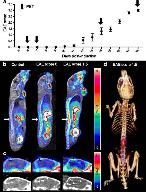
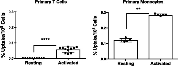
Methods: Experimental autoimmune encephalomyelitis (EAE) was induced in C57BL/6 mice by subcutaneous injection of myelin oligodendrocyte glycoprotein (MOG35-55) peptide in complete Freund's adjuvant (CFA) followed by pertussis toxin. Control mice received CFA emulsion and pertussis toxin without MOG peptide, while a separate cohort of naïve mice received no treatment. PET studies were performed to investigate the kinetics and distribution of [18F]FSPG in naïve, control, pre-symptomatic, and symptomatic EAE mice, compared to 18F-fluorodeoxyglucose ([18F]FDG). After final PET scans, each mouse was perfused and radioactivity in dissected tissues was measured using a gamma counter. Central nervous system (CNS) tissues were further analyzed using ex vivo autoradiography or western blot. [18F]FSPG uptake in human monocytes, and T cells pre- and post-activation was investigated in vitro.

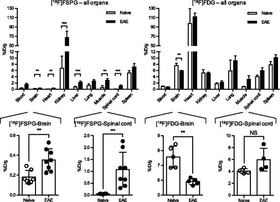
Results: [18F]FSPG was found to be more sensitive than [18F]FDG at detecting pathological changes in the spinal cord and brain of EAE mice. Even before clinical signs of disease, a small but significant increase in [18F]FSPG signal was observed in the spinal cord of EAE mice compared to controls. This increase in PET signal became more pronounced in symptomatic EAE mice and was confirmed by ex vivo biodistribution and autoradiography. Likewise, in the brain of symptomatic EAE mice, [18F]FSPG uptake was significantly higher than controls, with the largest changes observed in the cerebellum. Western blot analyses of CNS tissues revealed a significant correlation between light chain of xc- (xCT) protein levels, the subunit of xc- credited with its transporter activity, and [18F]FSPG-PET signal. In vitro [18F]FSPG uptake studies suggest that both activated monocytes and T cells contribute to the observed in vivo PET signal.

Conclusion: These data highlight the promise of [18F]FSPG-PET as a technique to provide insights into neuroimmune interactions in MS and the in vivo role of xc- in the development and progression of this disease, thus warranting further investigation.

Keywords: EAE mice; FSPG; Multiple sclerosis; PET; Xc-.


Conflict of interest statement

Ethics approval

All procedures performed on animals were approved by Stanford University’s Institutional Animal Care and Use Committee and were within the guidelines of humane care of laboratory animals (Protocol #20128).

Consent for publication

Not applicable.

Competing interests

The authors declare that they have no competing interests.

Publisher’s Note

Springer Nature remains neutral with regard to jurisdictional claims in published maps and institutional affiliations.

Figures

Fig. 1
Fig. 1
Ex vivo biodistribution of [18F]FSPG and [18F]FDG in EAE and naive mice. Ex vivo data was obtained 110 min after radiotracer injection (n = 5–9, mean %ID/g ± SD) with magnified views of uptake in brain and spinal cord. EAE scores: 2.6 ± 0.9 and 3.2 ± 0.3 for [18F]FSPG and [18F]FDG groups respectively. ***p 0.0001–0.001, **p 0.001–0.01
Fig. 2
Fig. 2
Elevated [18F]FSPG-PET signal in EAE mice at early stages of disease. a Disease scores of mice induced with EAE over 18 days. Arrows indicate PET/CT imaging time points and scores are represented as mean score ± SEM. b Representative sagittal PET/CT images of control, pre-symptomatic EAE (score 0), and EAE mice with early stage disease symptoms (score 1.5). Arrows indicate spinal cord uptake. Bl—bladder, Br—brain, H—Haderian glands, I—immunization site, K—kidneys, S—salivary glands. c Representative sagittal PET/CT images of identical brain sections of control, pre-symptomatic EAE (score 0), and symptomatic EAE (score 1.5) mice. Cb—cerebellum, HC—hippocampus, Mb—midbrain, Med—medulla. Dotted lines highlight region containing possible meningeal vessel(s). d Representative PET/CT volume rendering technique (VRT) image of EAE mouse (score 1.5). PET signal outside spinal column and skull was removed for better visualization of [18F]FSPG uptake in areas of interest
Fig. 3
Fig. 3
Quantification of [18F]FSPG-PET signal in CNS tissues of pre-symptomatic and symptomatic EAE versus control mice. Static PET imaging was performed 75 min post-injection of radiotracer (n = 6–10) and signal was quantified in the a whole spinal cord, b cervical/thoracic spinal cord, c lumbar spinal cord, and d whole brain. ***p 0.0001–0.001, **p 0.001–0.01, *p 0.01–0.05, NS—non-significant
Fig. 4
Fig. 4
Significant correlation between xCT protein levels in CNS tissues and in vivo PET signal. a Representative Western blot with actin and NaK-ATPase as loading controls (Ctrl). Correlation between xCT protein levels (determined by densitometric Western analysis) and [18F]FSPG uptake (determined by PET quantification) in the b cervical/thoracic spinal cord (SC), c lumbar SC, and d brain. ***p 0.0001–0.001, **p 0.001–0.01, *p 0.01–0.05
Fig. 5
Fig. 5
[18F]FSPG uptake in resting and activated primary human immune cells. In vitro accumulation of [18F]FSPG in isolated primary human T cells and monocytes (n = 5–11, mean %ID/g ± SD). ****p < 0.0001, **p < 0.01

Similar articles

  • In vivo imaging of system xc- as a novel approach to monitor multiple sclerosis.
    Martín A, Vázquez-Villoldo N, Gómez-Vallejo V, Padro D, Soria FN, Szczupak B, Plaza-García S, Arrieta A, Reese T, Llop J, Domercq M, Matute C. Martín A, et al. Eur J Nucl Med Mol Imaging. 2016 Jun;43(6):1124-38. doi: 10.1007/s00259-015-3275-3. Epub 2015 Dec 10. Eur J Nucl Med Mol Imaging. 2016. PMID: 26659901
  • Imaging B Cells in a Mouse Model of Multiple Sclerosis Using 64Cu-Rituximab PET.
    James ML, Hoehne A, Mayer AT, Lechtenberg K, Moreno M, Gowrishankar G, Ilovich O, Natarajan A, Johnson EM, Nguyen J, Quach L, Han M, Buckwalter M, Chandra S, Gambhir SS. James ML, et al. J Nucl Med. 2017 Nov;58(11):1845-1851. doi: 10.2967/jnumed.117.189597. Epub 2017 Jul 7. J Nucl Med. 2017. PMID: 28687602 Free PMC article.
  • 18F-VC701-PET and MRI in the in vivo neuroinflammation assessment of a mouse model of multiple sclerosis.
    Belloli S, Zanotti L, Murtaj V, Mazzon C, Di Grigoli G, Monterisi C, Masiello V, Iaccarino L, Cappelli A, Poliani PL, Politi LS, Moresco RM. Belloli S, et al. J Neuroinflammation. 2018 Feb 5;15(1):33. doi: 10.1186/s12974-017-1044-x. J Neuroinflammation. 2018. PMID: 29402285 Free PMC article.
  • (4S)-4-(3-[18F]Fluoropropyl)-l-glutamate.
    Chopra A. Chopra A. 2013 Mar 20 [updated 2013 May 23]. In: Molecular Imaging and Contrast Agent Database (MICAD) [Internet]. Bethesda (MD): National Center for Biotechnology Information (US); 2004–2013. 2013 Mar 20 [updated 2013 May 23]. In: Molecular Imaging and Contrast Agent Database (MICAD) [Internet]. Bethesda (MD): National Center for Biotechnology Information (US); 2004–2013. PMID: 23700646 Free Books & Documents. Review.
  • Glutamate, T cells and multiple sclerosis.
    Levite M. Levite M. J Neural Transm (Vienna). 2017 Jul;124(7):775-798. doi: 10.1007/s00702-016-1661-z. Epub 2017 Feb 24. J Neural Transm (Vienna). 2017. PMID: 28236206 Review.

Cited by

References

    1. Stadelmann C. Multiple sclerosis as a neurodegenerative disease: pathology, mechanisms and therapeutic implications. Curr Opin Neurol. 2011;24:224–229. doi: 10.1097/WCO.0b013e328346056f. - DOI - PubMed
    1. Gilgun-Sherki Y, Melamed E, Offen D. The role of oxidative stress in the pathogenesis of multiple sclerosis: the need for effective antioxidant therapy. J Neurol. 2004;251:261–268. doi: 10.1007/s00415-004-0348-9. - DOI - PubMed
    1. Conrad M, Sato H. The oxidative stress-inducible cystine/glutamate antiporter, system x c-: cystine supplier and beyond. Amino Acids. 2012;42:231–246. doi: 10.1007/s00726-011-0867-5. - DOI - PubMed
    1. Domercq M, Sánchez-Gómez MV, Sherwin C, Etxebarria E, Fern R, Matute C. System xc- and glutamate transporter inhibition mediates microglial toxicity to oligodendrocytes. J Immunol. 2007;178:6549–6556. doi: 10.4049/jimmunol.178.10.6549. - DOI - PubMed
    1. Kostic M, Zivkovic N, Stojanovic I. Multiple sclerosis and glutamate excitotoxicity. Rev Neurosci. 2013;24:71–88. doi: 10.1515/revneuro-2012-0062. - DOI - PubMed

MeSH terms

LinkOut - more resources