. 2018 Feb 27;71(8):875-886.
doi: 10.1016/j.jacc.2017.12.037.

Tet2-Mediated Clonal Hematopoiesis Accelerates Heart Failure Through a Mechanism Involving the IL-1β/NLRP3 Inflammasome

Affiliations

Tet2-Mediated Clonal Hematopoiesis Accelerates Heart Failure Through a Mechanism Involving the IL-1β/NLRP3 Inflammasome

Soichi Sano et al. J Am Coll Cardiol. .

Abstract

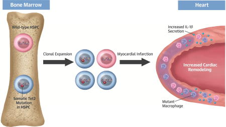
Background: Recent studies have shown that hematopoietic stem cells can undergo clonal expansion secondary to somatic mutations in leukemia-related genes, thus leading to an age-dependent accumulation of mutant leukocytes in the blood. This somatic mutation-related clonal hematopoiesis is common in healthy older individuals, but it has been associated with an increased incidence of future cardiovascular disease. The epigenetic regulator TET2 is frequently mutated in blood cells of individuals exhibiting clonal hematopoiesis.

Objectives: This study investigated whether Tet2 mutations within hematopoietic cells can contribute to heart failure in 2 models of cardiac injury.

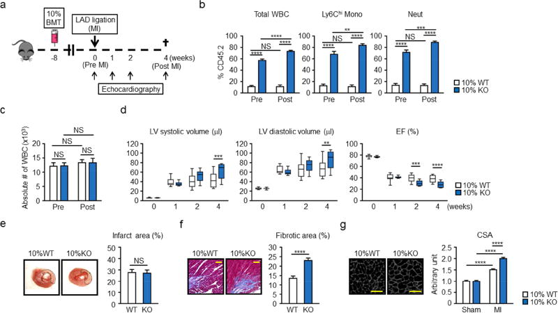
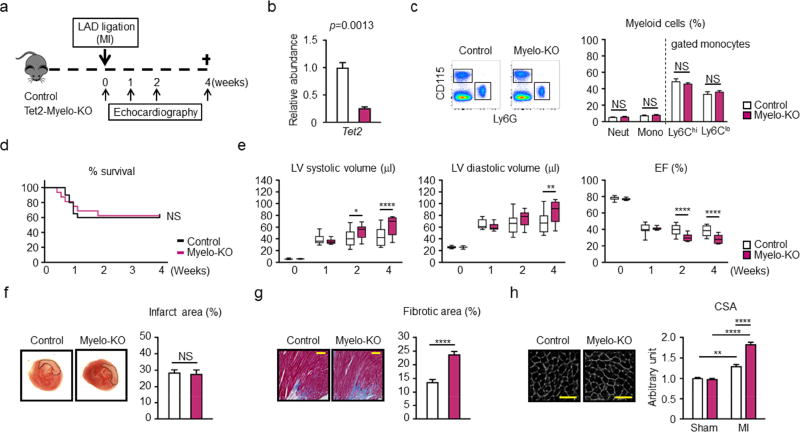
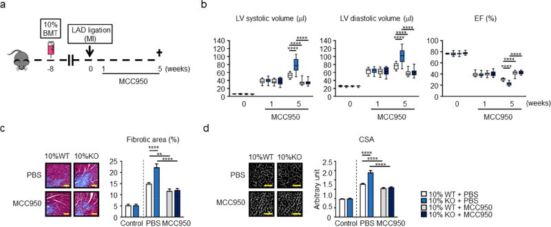
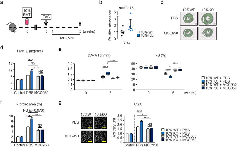
Methods: Heart failure was induced in mice by pressure overload, achieved by transverse aortic constriction or chronic ischemia induced by the permanent ligation of the left anterior descending artery. Competitive bone marrow transplantation strategies with Tet2-deficient cells were used to mimic TET2 mutation-driven clonal hematopoiesis. Alternatively, Tet2 was specifically ablated in myeloid cells using Cre recombinase expressed from the LysM promoter.

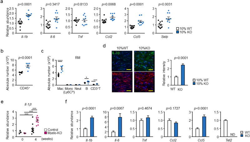
Results: In both experimental heart failure models, hematopoietic or myeloid Tet2 deficiency worsened cardiac remodeling and function, in parallel with increased interleukin-1beta (IL-1β) expression. Treatment with a selective NLRP3 inflammasome inhibitor protected against the development of heart failure and eliminated the differences in cardiac parameters between Tet2-deficient and wild-type mice.

Conclusions: Tet2 deficiency in hematopoietic cells is associated with greater cardiac dysfunction in murine models of heart failure as a result of elevated IL-1β signaling. These data suggest that individuals with TET2-mediated clonal hematopoiesis may be at greater risk of developing heart failure and respond better to IL-1β-NLRP3 inflammasome inhibition.

Keywords: 10-11 translocation 2; clonal hematopoiesis; heart failure; interleukin-1beta; myocardial infarction; pressure overload hypertrophy.


Figures

Figure 1
Figure 1. Hematopoietic Tet2-KO mice show greater post-infarction remodeling
a. Scheme of the experimental study. Mice underwent partial (10%) bone marrow reconstitution with Tet2-deficient cells (10% KO-BMT) or wild type cells (10% WT-BMT) following lethal irradiation. After 8 weeks of recovery, mice underwent permanent LAD ligation. Echocardiography was performed at the indicated time points. b. Tet2-KO bone marrow cells (Cd45.2+) display a competitive advantage over wild type competitor cells (Cd45.1+) in their ability to expand into multiple blood cell lineages in vivo. Peripheral blood was obtained 8 weeks and 12 weeks after BMT (before (Pre) and 4 weeks after (Post) MI, respectively) from 10% WT-BMT (n=11) mice and 10% KO-BMT mice (n=10). Statistical analysis was evaluated by evaluated by two-way ANOVA with Tukey’s multiple comparison tests. c. Absolute numbers of WBC before (Pre) and 4 weeks after (Post) LAD ligation of 10% KO-BMT mice (n=10) and 10% WT-BMT mice (n=11). Statistical analysis was evaluated by evaluated by two-way ANOVA with Tukey’s multiple comparison tests. d. Echocardiographic analysis shows that 10% KO-BMT mice (n=10) display worsening cardiac remodeling after LAD ligation compared to 10% WT-BMT mice (n=11). Statistical analysis was evaluated by two-way repeated measure ANOVA with Sidak’s multiple comparison tests. e. Representative images and analysis of infarct size in myocardial tissue sections from 10% WT-BMT (n=3) mice and 10% KO-BMT mice (n=3) stained with TTC 2 days after LAD ligation, showing there is no statistical significance between both groups. Hearts were sliced at 2 mm below from the ligation site. Statistical analysis was evaluated by two-tailed unpaired Student’s t test. f. Representative images and analysis of fibrosis in marginal zone of myocardial tissue sections from 10% WT-BMT (n=7) mice and 10% KO-BMT mice (n=7) stained with Masson’s Trichrome dye at 4 weeks after ligation, showing worsening fibrosis in 10% KO-BMT mice. The percentage of the fibrotic area was calculated with the image-J software. Statistical analysis was evaluated by two-tailed unpaired Student’s t test. Scale bars indicate 100 µm. g. Representative images and analysis of WGA staining of the heart sections from hearts 10% WT-BMT (n=7) mice and 10% KO-BMT mice (n=7) isolated at 4 weeks after LAD ligation. Staining shows that the non-infarcted, remote area of the heart displays greater hypertrophy of the cardiac myocytes. Statistical analysis was evaluated by two-way ANOVA with Tukey’s multiple comparison test. Scale bars indicate 50 µm. **p<0.01, ***p<0.001, ****p<0.0001, NS: not significant. WT: wild type, KO: knockout, BMT: bone marrow transfer, LAD: left anterior ascending artery. WBC: white blood cells, Mono: monocytes, Neut: neutrophils, LV: left ventricle, EF: ejection fraction, TTC: 2,3,5-triphenyl-tetrazolium chloride, CSA: cross-sectional area of myocytes.
Figure 2
Figure 2. Conditional myeloid Tet2-deficiency in mice leads worse cardiac remodeling in hearts subjected to LAD ligation
a. Scheme of the study. Control and conditional myeloid Tet2-knockout (Myelo-KO) mice underwent LAD ligation. Mice underwent permanent LAD ligation, and echocardiography was performed at the indicated time points. b. The efficiency of Tet2 ablation was analyzed by qPCR in BMDM at 7 days after in vitro differentiation from conditional Tet2-Myelo-KO mice and control mice (3 mice per genotype). Two-tailed Student’s t test was performed for statistical analysis. c. Flow cytometry representative data and analysis of peripheral blood from Tet2-Myelo-KO mice (n=6) and control mice (n=6) to show there are no detectable changes in myeloid populations. Statistical significance of difference was evaluated by multiple t test. d. Mice survival curve after LAD ligation. The mortality of the conditional KO mice and control mice after surgery was 37.5% and 40.0%, respectively. Log-rank test was used for statistical analysis (n= 20 for control mice and n= 16 for conditional Tet2-KO mice). e. Echocardiographic evaluation shows that surviving mice with conditional Tet2 ablation in myeloid cells (n=10) display worsening cardiac remodeling after LAD ligation surgery compared to control mice (n=12). Statistical analysis was evaluated by two-way repeated measure ANOVA with Sidak’s multiple comparison tests. f. Representative images and analysis of infarct size in myocardial tissue sections from conditional KO mice (n=3) and control (n=3) mice stained with TTC 2 days after LAD ligation. Hearts were sliced at 2 mm below from the ligation site, showing that there was no difference in initial infarct size. Statistical analysis was evaluated by Mann-Whitney U test. g. Representative images and analysis of fibrosis in the marginal zone of myocardial tissue sections from conditional KO mice (n=6) and control (n=6) mice stained with Masson’s Trichrome dye at 4 weeks after ligation, showing worsening fibrosis in conditional KO mice. The percentage of the fibrotic area was calculated with the image-J software. Statistical analysis was evaluated by two-tailed unpaired Student’s t test. Scale bars indicate 100 µm. h. WGA staining of the heart sections from hearts control (n=6) mice and conditional KO mice (n=6) isolated at 4 weeks after LAD ligation. Analysis of CSA shows that the non-infarcted, remote area of the heart of conditional KO mice display greater hypertrophy of the cardiac myocytes. Statistical analysis was evaluated by two-way ANOVA followed with Tukey’s multiple comparison tests. Scale bars indicate 50 µm. *p<0.05, **p<0.01, ****p<0.0001, NS: not significant. WT: wild type, KO: knockout, LAD: left anterior ascending artery, qPCR: quantitative polymerase chain reaction, BMDM: bone marrow-derived macrophages, LAD: left anterior ascending artery, Mono: monocytes, Neut: neutrophils, LV: left ventricle, EF: ejection fraction, TTC: 2,3,5-triphenyl-tetrazolium chloride, WGA: wheat germ agglutinin, CSA: cross-sectional area of myocytes.
Figure 3
Figure 3. Effect of Tet2-deficient hematopoietic cells on the expression of pro-inflammatory cytokines and chemokines in the remodeling heart tissue
a. Analysis of transcript expression in the non-infarcted marginal zone obtained from 10% KO-BMT mice (n=10) and 10% WT-BMT (n=11) mice. Gene expression was analyzed by qPCR analysis. Statistical significance was evaluated by two-tailed unpaired Student’s t tests with Welch’s Correction when variance was unequal or by Mann Whitney U tests for data which failed to pass the Shapiro-Wilk normality test. b. Flow cytometry analysis of cardiac remote area from 10% KO-BMT mice (n=7) and 10% WT-BMT (n=7) mice to show the absolute number of total CD45+ immune cells are increased in the myocardial tissue from 10% KO-BMT mice. Data is expressed as number of cells per 100 mg wet weight. Statistical analysis was evaluated by two-tailed unpaired Student’s t test. c. Flow cytometry analysis of cardiac remote area from 10% KO-BMT mice (n=7) and 10% WT-BMT (n=7) mice to show the absolute number of each immune cell populations. Statistical significance of difference was evaluated by multiple t tests. d. IL-1β immunofluorescence staining in Mac3-positive macrophage-enriched marginal zone of 10% KO-BMT (n=5) mice and 10% WT-BMT mice (n=5) showing IL-1β signal is higher in 10% KO-BMT mice. Scale bars = 20 µm. Images were quantified for integrated fluorescence intensity with Image J software. Statistical analysis was performed by two-tailed unpaired Student’s t tests. e. Remote area samples were obtained from conditional myeloid-specific KO mice and control mice, and gene expression was analyzed by qPCR at the indicated time points (n=3 for sham and n = 10 at 4 weeks after LAD ligation, per genotype). Statistical significance was evaluated by two-way ANOVA with Tukey’s multiple comparison tests. f. Bone marrow-derived macrophages 2 days after in vitro differentiation obtained from Tet2-null mice (n=6) and wild type (n=7) were obtained in vitro and gene expression was analyzed by qPCR analysis. Statistical significance of difference was evaluated by two-tailed unpaired Student’s t tests with Welch’s Correction when variance was unequal or by Mann Whitney U tests for data which failed to pass the Shapiro-Wilk normality test. *p<0.05, **p<0.01, ****p<0.0001. WT: wild type, KO: knockout, BMT: bone marrow transfer, LAD: left anterior ascending artery, qPCR: quantitative polymerase chain reaction, RM: remote area, Mac: macrophages, Mono: monocytes, Neut: neutrophils, B: B cells, T: T cells, ND: not detected.
Figure 4
Figure 4. Inflammasome inhibition reverses post-infarction remodeling associated with hematopoietic Tet2-deficiency
a. Scheme of the experimental study. Mice underwent partial (10%) bone marrow reconstitution with Tet2-deficient cells (10% KO-BMT mice) or wild type cells (10% WT-BMT mice) following lethal irradiation. After 8 weeks of recovery, mice underwent permanent LAD ligation. 1 week after LAD ligation, MCC950 and PBS was continuously infused with osmotic pumps for 4 weeks. Echocardiography was performed at the indicated time points. b. Echocardiographic analysis reveals that treatment with the NLRP3 inflammasome inhibitor MCC950 protects against adverse cardiac remodeling in 10% KO-BMT and 10% WT-BMT mice, and eliminates the differences in cardiac parameters between Tet2-deficient and WT conditions at the post-LAD ligation time point of 5 weeks. Sample sizes were n = 12 for 10% WT-BMT with PBS, n=12 for 10% KO-BMT with PBS, n = 14 for 10% WT-BMT with MCC950, n = 14 for 10% KO-BMT with MCC950. Statistical significance was evaluated by 2-way repeated measure ANOVA with Tukey’s multiple comparison tests. c. Representative images and analysis of fibrosis in marginal zone of myocardial tissue sections stained with Masson’s Trichrome dye at 5 weeks after ligation. MCC950 inhibits the development of cardiac fibrosis after LAD ligation in mice reconstituted with Tet2-KO or WT bone marrow, and eliminates the differences in cell size between Tet2-deficient and WT genotypes. Statistical significances of differences among groups of 10% WT/10%KO with PBS or MCC950 were evaluated by two-way ANOVA with Tukey’s multiple comparison tests. Scale bars indicate 100 µm. d. Representative images and analysis of WGA staining of the heart sections of hearts at 5 weeks after LAD ligation. MCC950 inhibits the development of cardiac myocyte hypertrophy after LAD ligation in mice reconstituted with Tet2-KO or WT bone marrow, and eliminates the differences in cell size between Tet2-deficient and WT genotypes. For c and d, sham-operated mice without any pump infusion were used as control (n=3 per genotype). Sample sizes were n=6 for 10% WT-BMT with PBS, n=6 for 10% KO-BMT with PBS, n=8 for 10% WT-BMT with MCC950, n=8 for 10% KO-BMT with MCC950. Statistical significances of differences among groups of 10% WT/10%KO with PBS or MCC950 were evaluated by two-way ANOVA with Tukey’s multiple comparison tests. Scale bars indicate 50 µm. **p<0.01, ***p<0.001, ****p<0.0001. WT: wild type, KO: knockout, BMT: bone marrow transfer, LAD: left anterior ascending artery, PBS: phosphate-buffered saline, WGA: wheat germ agglutinin, CSA: cross-sectional area of myocytes.
Figure 5
Figure 5. Inflammasome inhibition reverses pressure overload-induced cardiac remodeling associated with hematopoietic Tet2-deficiency
a. Scheme of the experimental study. Mice underwent partial (10%) bone marrow reconstitution with Tet2-deficient cells (10% KO-BMT mice) or WT cells (10% WT-BMT) following lethal irradiation. After 8 weeks of recovery, mice underwent permanent TAC surgery to produce pressure overload on the heart. MCC950 or PBS were infused from 1 week after TAC. b. IL-1β transcripts were determined in heart samples from 10% WT-BMT (n=7) mice and 10% KO-BMT mice (n=7) after pressure overload were by qPCR analysis. Statistical significance was evaluated by Mann-Whitney U test. c. Representative images of Picrosirius red staining to show the heart from 10% KO-BMT mice is larger compared to the heart from 10% WT-BMT mice 5 weeks after TAC. Scale bar indicates 1 mm. d. HW adjusted by TL, showing that MCC950 ameliorates the increase of cardiac mass after pressure overload in both strains of mice and eliminates the differences in these parameters between the Tet2-deficient and WT conditions (n=7 for TAC with PBS and n=8 for TAC with MCC950 per genotype). Sham operated mice without any infusion were used as control (n=3 per genotype). Statistical significances of differences among groups of 10% WT/10%KO with PBS or MCC950 were evaluated by two-way ANOVA with Tukey’s multiple comparison tests. e. Echocardiographic analysis shows that infusion with MCC950 protects against adverse cardiac remodeling in mice reconstituted with Tet2-KO and wild-type bone marrow, and eliminates the differences in cardiac parameters between Tet2-deficient and WT genotypes at the 5 weeks after TAC surgery. Echocardiography was performed at the indicated time points. Sample sizes were n=7 for 10% WT-BMT with PBS, n=7 for 10% KO-BMT with PBS, n=8 for 10% WT-BMT with MCC950, n=8 for 10% KO-BMT with MCC950. Statistical significance of difference was evaluated by two-way repeated measure ANOVA with Tukey’s multiple comparison tests. f. Quantitative analysis of cardiac sections stained with Picro sirius red as presented in Fig. 5c. shows that mice reconstituted with Tet2-knockout bone marrow exhibit greater cardiac fibrosis after pressure overload that can be reversed by treatment with MCC950. The MCC950 treatment eliminates the difference in this parameter between the Tet2-deficient and WT conditions. Sample sizes were n=7 for 10% WT-BMT with PBS, n=7 for 10% KO-BMT with PBS, n=8 for 10% WT-BMT with MCC950, n=8 for 10% KO-BMT with MCC950. Sham mice without any infusion were used as control (n=5 per genotype). Statistical significances of differences among groups of 10% WT/10%KO with PBS or MCC950 were evaluated by two-way ANOVA with Tukey’s multiple comparison tests. g. Representative images and measurement of CSA stained with WGA shows that MCC950 inhibits hypertrophy after pressure overload both in wild type and hematopoietic Tet2-KO mice and eliminates the difference in parameters between the Tet2-deficient and WT conditions. Sample sizes were n=7 for 10% WT-BMT with PBS, n=7 for 10% KO-BMT with PBS, n=8 for 10% WT-BMT with MCC950, n=8 for 10% KO-BMT with MCC950. Sham mice without any infusion were used as control (n=5 per genotype). Statistical significances of differences among groups of 10% WT/10%KO with PBS or MCC950 were evaluated by two-way ANOVA with Tukey’s multiple comparison tests. *p<0.05, ****p<0.0001; NS: not significant. WT: wild type, KO: knockout, BMT: bone marrow transfer, TAC: transverse aortic constriction, PBS: phosphate-buffered saline, qPCR: quantitative polymerase chain reaction, HW: heart weight, TL: tibia length, WGA: wheat germ agglutinin, WGA: wheat germ agglutinin, CSA: cross-sectional area of myocytes.
Central Illustration
Central Illustration. Clonal Hematopoiesis Promotes Heart Failure
Somatic Tet2 mutations within hematopoietic stem and progenitor cells (HSPC) will lead to their clonal amplification. These HSPC give rise to myeloid cell progeny that promote cardiac remodeling through excessive production of IL-1β.

Comment in

References

    1. de Freitas EV, Batlouni M, Gamarsky R. Heart failure in the elderly. J Geriatr Cardiol. 2012;9:101–7. - PMC - PubMed
    1. Belsky DW, Caspi A, Houts R, et al. Quantification of biological aging in young adults. Proc Natl Acad Sci U S A. 2015;112:E4104–10. - PMC - PubMed
    1. Vigen R, Maddox TM, Allen LA. Aging of the United States population: impact on heart failure. Curr Heart Fail Rep. 2012;9:369–74. - PMC - PubMed
    1. Vijg J. Somatic mutations, genome mosaicism, cancer and aging. Curr Opin Genet Dev. 2014;26:141–9. - PMC - PubMed
    1. Shlush LI, Zandi S, Itzkovitz S, Schuh AC. Aging, clonal hematopoiesis and preleukemia: not just bad luck? Int J Hematol. 2015;102:513–22. - PubMed

Publication types

MeSH terms