

HARMless: Transient Cortical and Sulcal Hyperintensity on Gadolinium-Enhanced FLAIR after Elective Endovascular Coiling of Intracranial Aneurysms
- PMID: 29472303
- PMCID: PMC7410778
- DOI: 10.3174/ajnr.A5561
HARMless: Transient Cortical and Sulcal Hyperintensity on Gadolinium-Enhanced FLAIR after Elective Endovascular Coiling of Intracranial Aneurysms
Abstract
Background and purpose: Cortical and sulcal hyperintensity on gadolinium-enhanced FLAIR has been increasingly recognized after iodinated contrast medium exposure during angiographic procedures. The goal of this study was to assess the relationship of cortical and sulcal hyperintensity on gadolinium-enhanced FLAIR against various variables in patients following elective endovascular treatment of intracranial aneurysms.
Materials and methods: We performed a retrospective review of 58 patients with 62 MR imaging studies performed within 72 hours following endovascular treatment of intracranial aneurysms. Patient demographics, aneurysm location, and vascular territory distribution of cortical and sulcal hyperintensity on gadolinium-enhanced FLAIR were documented. Analysis of cortical and sulcal hyperintensity on gadolinium-enhanced FLAIR with iodinated contrast medium volume, procedural duration, number of angiographic runs, and DWI lesions was performed.
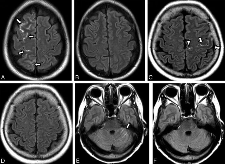
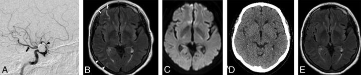
Results: Cortical and sulcal hyperintensity on gadolinium-enhanced FLAIR was found in 32/62 (51.61%) post-endovascular treatment MR imaging studies, with complete resolution of findings in all patients on the available follow-up studies (27/27). Angiographic iodinated contrast medium injection and arterial anatomy matched the vascular distribution of cortical and sulcal hyperintensity on gadolinium-enhanced FLAIR. No significant association was found between cortical and sulcal hyperintensity on gadolinium-enhanced FLAIR with iodinated contrast medium volume (P = .56 value) and the presence of DWI lesions (P = .68). However, a significant association was found with procedural time (P = .001) and the number of angiographic runs (P = .019). No adverse clinical outcomes were documented.
Conclusions: Cortical and sulcal hyperintensity on gadolinium-enhanced FLAIR is a transient observation in the arterial territory exposed to iodinated contrast medium during endovascular treatment of intracranial aneurysms. Cortical and sulcal hyperintensity on gadolinium-enhanced FLAIR is significantly associated with procedural time, and the frequency of angiographic runs suggesting a potential technical influence on the breakdown of the BBB, but no reported adverse clinical outcome or association with both iodinated contrast medium volume and DWI lesions was found. Recognition of cortical and sulcal hyperintensity on gadolinium-enhanced FLAIR as a benign incidental finding is vital to avoid unnecessary investigation.
© 2018 by American Journal of Neuroradiology.
Figures
References
-
- Ogami R, Nakahara T, Hamasaki O, et al. Cerebrospinal fluid enhancement on fluid attenuated inversion recovery images after carotid artery stenting with neuroprotective balloon occlusions: hemodynamic instability and blood-brain barrier disruption. Cardiovasc Intervent Radiol 2011;34:936–41 10.1007/s00270-010-0035-4 - DOI - PubMed
MeSH terms
Substances
LinkOut - more resources
Full Text Sources
Other Literature Sources
Medical