. 2017:82:279-291.
doi: 10.1101/sqb.2017.82.034439. Epub 2018 Feb 22.

Dynamic Control of Chromosome Topology and Gene Expression by a Chromatin Modification

Affiliations

Dynamic Control of Chromosome Topology and Gene Expression by a Chromatin Modification

Qian Bian et al. Cold Spring Harb Symp Quant Biol. 2017.

Abstract

The function of chromatin modification in establishing higher-order chromosome structure during gene regulation has been elusive. We dissected the machinery and mechanism underlying the enrichment of histone modification H4K20me1 on hermaphrodite X chromosomes during Caenorhabditis elegans dosage compensation and discovered a key role for H4K20me1 in regulating X-chromosome topology and chromosome-wide gene expression. Structural and functional analysis of the dosage compensation complex (DCC) subunit DPY-21 revealed a novel Jumonji C demethylase subfamily that converts H4K20me2 to H4K20me1 in worms and mammals. Inactivation of demethylase activity in vivo by genome editing eliminated H4K20me1 enrichment on X chromosomes of somatic cells, increased X-linked gene expression, reduced X-chromosome compaction, and disrupted X-chromosome conformation by diminishing the formation of topologically associated domains. H4K20me1 is also enriched on the inactive X of female mice, making our studies directly relevant to mammalian development. Unexpectedly, DPY-21 also associates specifically with autosomes of nematode germ cells in a DCC-independent manner to enrich H4K20me1 and trigger chromosome compaction. Thus, DPY-21 is an adaptable chromatin regulator. Its H4K20me2 demethylase activity can be harnessed during development for distinct biological functions by targeting it to diverse genomic locations through different mechanisms. In both somatic cells and germ cells, H4K20me1 enrichment modulates three-dimensional chromosome architecture, demonstrating the direct link between chromatin modification and higher-order chromosome structure.


Figures

Figure 1
Figure 1
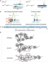
Overview of dosage compensation in Caenorhabditis elegans. (A) In XX hermaphrodites, a dosage compensation complex (DCC) binds to both X chromosomes to reduce gene expression by half, thereby equalizing expression with that from the single male X. The product of the XX-specific gene sdc-2 triggers assembly of the DCC onto X. In XO males, xol-1, the male-specific regulator of sex determination and dosage compensation, represses sdc-2, thereby preventing the DCC from binding to the male X. (B) The DCC compared with condensin I of other eukaryotes. The DCC condensin subunits (MIX-1, DPY-27, DPY-26, DPY-28, and CAPG-1) are color matched to their condensin I homologs (Csankovszki et al. 2009; Mets and Meyer 2009; Meyer 2010). All DCC condensin subunits except DPY-27 also function in other condensins that act in C. elegans mitosis and meiosis. The DPY-27 paralog SMC-4 (not shown) replaces DPY-27 in mitotic and meiotic condensins. The DCC likely arose by duplicating the gene encoding SMC-4 and modifying it to create DPY-27 for a specific role in gene expression (Hagstrom et al. 2002). In addition to condensin subunits, the DCC also includes a novel XX-specific protein with a large coiled-coil domain (SDC-2) (Dawes et al. 1999) that triggers assembly of the DCC onto X chromosomes. Two DCC subunits aid SDC-2 in recruiting the complex to X, SDC-3 (a zinc finger protein) and DPY-30 (a subunit of the MLL/COMPASS H3K4me3 methyltransferase complex) (Klein and Meyer 1993; Hsu et al. 1995; Davis and Meyer 1997; Pferdehirt et al. 2011). Two subunits, SDC-1 (a zinc finger protein) and DPY-21, are required for DCC activity but not assembly (Nonet and Meyer 1991; Yonker and Meyer 2003). DPY-21 is a Jumonji C H4K20me2 demethylase described here and in (Brejc et al. 2017). (C) DCC recruitment sites across X chromosomes. The DCC recruitment elements on X (rex) were discovered by the combination of genome-wide approaches (ChIP-chip and ChIP-seq) to identify DCC-binding sites without regard to autonomous recruitment ability and a functional approach in vivo to assess DCC binding to sites detached from X (Jans et al. 2009; Crane et al. 2015). rex sites are distributed across X and confer X-chromosome specificity to dosage compensation. DCC binding to rex sites facilitates DCC spreading across X to sites that cannot bind the complex if detached from X (Pferdehirt et al. 2011). Several of the strongest rex sites (red) are essential for formation of topologically associated domains (TADs). (D) Cartoon model of TAD formation on a segment of X. (Top) The DCC remodels the topology of X into a hermaphrodite-specific conformation by forming TADs. DCC-dependent looping interactions between high-affinity rex sites located at TAD boundaries direct TAD formation (Crane et al. 2015). (Middle) Deletion of the high-affinity site rex-47 located at a DCC-dependent TAD boundary eliminates boundary formation (Crane et al. 2015). (Bottom) Severe disruption of DCC binding by an sdc-2 mutation eliminates formation of all DCC-dependent TADs on X (Crane et al. 2015).
Figure 2
Figure 2
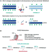
H4K20me1 enrichment on the repressed X chromosomes is a shared feature of diverse dosage compensation strategies. (A) Dosage-compensated X chromosomes of C. elegans hermaphrodites have dosage compensation complex (DCC)-dependent H4K20me1 enrichment. H4K20me1 enrichment on the inactive X chromosome of female mammals requires the long noncoding RNA XIST that triggers X inactivation. For neither strategy had the mechanism of H4K20me1 enrichment been determined. (B) H4K20 methylation controls myriad nuclear functions, but the mechanisms that regulate different H4K20 methylation states are not well understood. H4K20me2/me3 demethylases had not been identified.
Figure 3
Figure 3
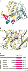
1.8 Å structure of the DPY-21 JmjC demethylase domain. (A) DPY-211210–1617 structure in complex with α-KG (black) and Fe2+ (orange) showing JmjC domain (yellow), JmjN (blue), β-hairpin (magenta), and mixed domain (green). (Adapted from Brejc et al. 2017.) (B) Active site of DPY-211210–1617 showing JmjC domain residues (yellow) in complex with Fe2+ (orange), α-KG (black), and water molecules (red). Facial triad residues H1452 and D1454 (red letters) were changed to alanines for in vitro and in vivo studies. The electron density, 2FoFc (mesh), contoured at 1.0 σ above the mean is shown for Fe2+, α-KG, and water molecules. (Adapted from Brejc et al. 2017.) (C) Evolutionary conservation of DPY-21 JmjC domain (magenta) in ROSBIN proteins across species.
Figure 4
Figure 4
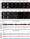
DPY-21 JmjC H4K20me2 demethylase enriches H4K20me1 on X in vivo. (A) Confocal images of an interphase nucleus from a 376-cell wild-type embryo (top) and an interphase nucleus from a 335-cell dpy-21(JmjC) mutant embryo (bottom) stained with DAPI and antibodies to DPY-21, dosage compensation complex (DCC) subunit SDC-3, and H4K20me1. The JmjC mutation does not affect binding of DPY-21 to X, but it does disrupt the H4K20me1 enrichment on X. (B) Metaphase nucleus from a 376-cell wild-type embryo (top) and metaphase nucleus from a 335-cell dpy-21(JmjC) mutant embryo (bottom) stained as in A. During mitosis, DPY-21 dissociates from X, but SDC-3 remains bound. The JmjC mutation does not affect the H4K20me1 level on mitotic chromosomes. (C) ChIP-seq profiles show spike-in-corrected H4K20me1 enrichment in representative regions of chromosome X and chromosome IV in wild-type, dpy-21(JmjC), and dpy-21(null) mutant embryos. X enrichment of H4K20me1 is lost in dpy-21 mutants, but H4K20me1 levels are unchanged on autosomes. (Based on data from Brejc et al. 2017.)
Figure 5
Figure 5
X-chromosome gene expression is elevated relative to autosomes in dpy-21(JmjC) mutants. Cumulative plots show the distribution of expression changes for genes on X and for each individual autosome in dpy-21(JmjC) mutant versus wild-type embryos as assayed by RNA-seq. The x-axis represents the log2 fold change in expression. X-chromosome gene expression is elevated compared to that of each autosome in dpy-21(y607 JmjC), dpy-21(y618 JmjC), dpy-21(e428 null) mutants (P < 2.2 × 10⁻¹⁶, one-sided Wilcoxon rank-sum test). (Based on data from Brejc et al. 2017.)
Figure 6
Figure 6
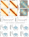
DPY-21 demethylase activity modulates X-chromosome topology. (A,B,D,E) Heat maps of Hi-C data binned at 50-kb resolution show chromatin interaction frequencies on chromosomes X and I in wild-type and dpy-21(JmjC) mutant embryos. These heat maps show results from one of two replicates. Heat maps combining both replicates are presented in Brejc et al. (2017), with the same conclusions. (C,F) Z-score difference heat maps of Hi-C data in A,B and D,E, respectively, binned at 50-kb resolution show chromatin interactions that increase (orange-red) and decrease (blue) on X (C) and I (F) in dpy-2(JmjC) mutant versus wild-type embryos. (G,H) Insulation plots for chromosomes X or I of wild-type (gray) or dpy-21(JmjC) mutant (blue) embryos and insulation difference plots (red) from data in AE. Black bars, location of topologically associated domain (TAD) boundaries in wild-type embryos. Red dots, dosage compensation complex (DCC)-dependent boundaries greatly diminished or eliminated upon DCC depletion (Crane et al. 2015). Each has a high-affinity rex site. An insulation score reflects the cumulative interactions occurring across each interval. Minima denote areas of high insulation classified as TAD boundaries. The difference between insulation profiles of wild-type and dpy-21(JmjC) mutants reflects the change in boundary strength. An increase in insulation score at a TAD boundary means less insulation in the mutant, indicating a weakening of the boundary. DCC-dependent TAD boundaries on X were reduced in dpy-21(JmjC) mutants, but DCC-independent boundaries on X and autosomes were not significantly changed. (I) Three-dimensional profiles of average Hi-C interaction frequencies (Z-scores) in 50-kb bins around pairs of top 25 rex sites, DCC-dependent boundaries on X, and DCC-independent boundaries on X in wild-type or dpy-21(JmjC) mutant embryos. Profiles are centered at 0. Interactions between rex sites and DCC-dependent boundaries are reduced in demethylase mutants. (Adapted from Brejc et al. 2017.)
Figure 7
Figure 7
DPY-21 JmjC demethyase acts in germ cells to enrich H4K20me1 and compact autosomes. (A) Confocal images of metaphase chromosomes from the mitotic zones of dpy-21(5′-FLAG) and dpy-21(5′-FLAG, JmjC) gonads costained with H4K20me1 and FLAG antibodies. H4K20me1 is highly enriched on all mitotic chromosomes, but DPY-21 does not localize to mitotic chromosomes. Inactivation of the DPY-21 demethylase does not reduce H4K20me1 levels on mitotic chromosomes. Scale bar, 1 μm. (B) Pachytene nuclei from dpy-21(5-FLAG) and dpy-21(5-FLAG, JmjC) XX gonads stained with antibodies against H4K20me1 and the X-chromosome-specific marker HIM-8. H4K20me1 is selectively enriched on autosomes in pachytene nuclei in the dpy-21(5-Flag) strain but absent from X (top). H4K20me1 is absent from all chromosomes in pachytene nuclei of dpy-21(JmjC) mutant XX gonads (bottom). (C) Pachytene nuclei from dpy-21(5-Flag) and dpy-21(5-Flag, JmjC) XX gonads stained with antibodies against FLAG-tagged DPY-21 and HIM-8. DPY-21 specifically localizes to autosomes but not to X, consistent with its demethylation of only autosomes. Scale bars (B, C), 2 μm. (D) DPY-21 demethylase is required for full compaction of autosomes in germ cells. (Left) High-resolution image of a pachytene nucleus from a wild-type gonad stained with a FISH probe to chromosome I (blue) and antibodies to axis protein HTP-3 (green) and X-specific protein HIM-8 (red). The 3D traces of X and I (yellow) were used to straighten each chromosome. (Middle) Computationally straightened X and I from wild-type and dpy-21(JmjC) gonads are displayed horizontally. Chromosome tracing was performed on nuclei in the last quarter of pachytene. Average total axis length and standard error of the mean (SEM) are shown below each axis. (Right) Box plots show the ratios of I to X axis lengths in wild-type and dpy-21(JmjC) pachytene nuclei. The I to X ratios are significantly lower in wild-type versus dpy-21(JmjC) gonads (P = 1.7 × 10⁻⁸, two-sided Wilcoxon rank-sum test). (Adapted from Brejc et al. 2017.)
Figure 8
Figure 8
An H4K20me2 histone demethylase regulates 3D chromosome structure and gene expression by modulating the dynamic enrichment of H4K20me1. The 1.8 Å crystal structure and biochemical activity of DPY-21 revealed a new, highly conserved H4K20me2 JmjC demethylase subfamily that converts H4K20me2 to H4K20me1 in vitro. In somatic cells, DPY-21 binds to X chromosomes via the dosage compensation complex (DCC) and enriches H4K20me1 to repress gene expression. The H4K20me1 enrichment controls the higher-order structure of X chromosomes by facilitating compaction and topologically associated domain (TAD) formation. In germ cells, DPY-21 enriches HK20me1 on autosomes in a DCC-independent manner to promote chromosome compaction. (Adapted from Brejc et al. 2017.)

References

    1. Beck DB, Oda H, Shen SS, Reinberg D. PR-Set7 and H4K20me1: At the crossroads of genome integrity, cell cycle, chromosome condensation, and transcription. Genes Dev. 2012;26:325–337. - PMC - PubMed
    1. Blanchard DP, Georlette D, Antoszewski L, Botchan MR. Chromatin reader L(3)mbt requires the Myb-MuvB/DREAM transcriptional regulatory complex for chromosomal recruitment. Proc Natl Acad Sci. 2014;111:E4234–E4243. - PMC - PubMed
    1. Boettiger AN, Bintu B, Moffitt JR, Wang S, Beliveau BJ, Fudenberg G, Imakaev M, Mirny LA, Wu C-T, Zhuang X. Super-resolution imaging reveals distinct chromatin folding for different epigenetic states. Nature. 2016;529:418–422. - PMC - PubMed
    1. Brejc K, Bian Q, Uzawa S, Wheeler BS, Anderson EC, King DS, Kranzusch PJ, Preston CG, Meyer BJ. Dynamic control of X chromosome conformation and repression by a histone H4K20 demethylase. Cell. 2017;171:85–102 e123. - PMC - PubMed
    1. Chen Z, Zang J, Whetstine J, Hong X, Davrazou F, Kutateladze TG, Simpson M, Mao Q, Pan CH, Dai S, et al. Structural insights into histone demethylation by JMJD2 family members. Cell. 2006;125:691–702. - PubMed

LinkOut - more resources