. 2018 Feb 22;8(1):3481.
doi: 10.1038/s41598-018-21869-x.

Inhibitory modulation of cytochrome c oxidase activity with specific near-infrared light wavelengths attenuates brain ischemia/reperfusion injury

Affiliations

Inhibitory modulation of cytochrome c oxidase activity with specific near-infrared light wavelengths attenuates brain ischemia/reperfusion injury

Thomas H Sanderson et al. Sci Rep. .

Erratum in

Abstract

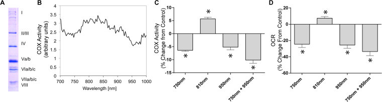
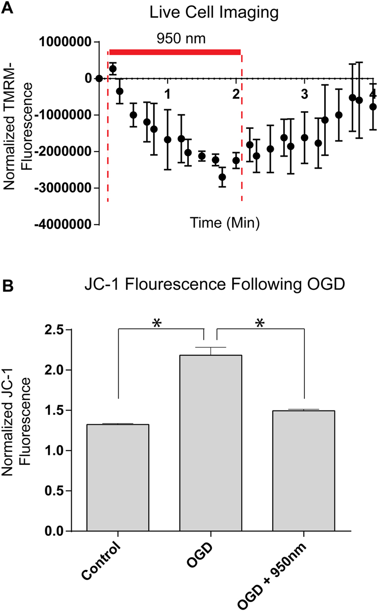
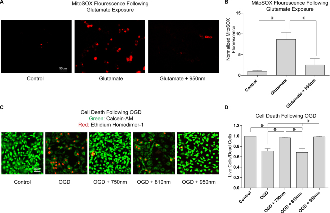
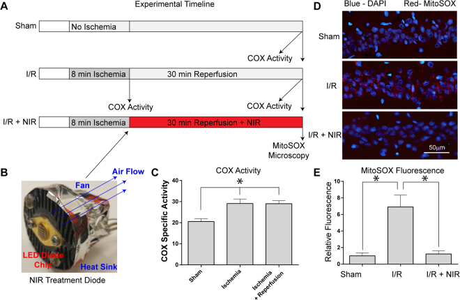
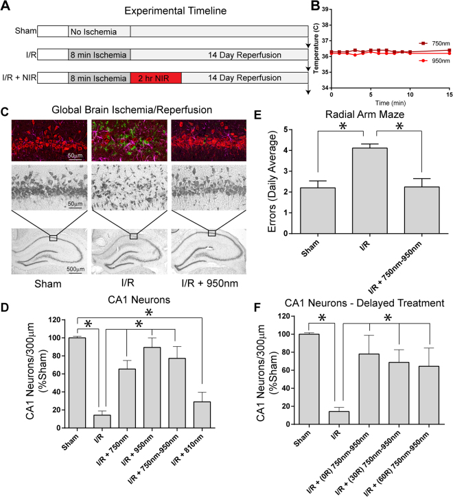
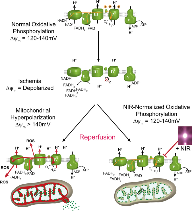
The interaction of light with biological tissue has been successfully utilized for multiple therapeutic purposes. Previous studies have suggested that near infrared light (NIR) enhances the activity of mitochondria by increasing cytochrome c oxidase (COX) activity, which we confirmed for 810 nm NIR. In contrast, scanning the NIR spectrum between 700 nm and 1000 nm revealed two NIR wavelengths (750 nm and 950 nm) that reduced the activity of isolated COX. COX-inhibitory wavelengths reduced mitochondrial respiration, reduced the mitochondrial membrane potential (ΔΨm), attenuated mitochondrial superoxide production, and attenuated neuronal death following oxygen glucose deprivation, whereas NIR that activates COX provided no benefit. We evaluated COX-inhibitory NIR as a potential therapy for cerebral reperfusion injury using a rat model of global brain ischemia. Untreated animals demonstrated an 86% loss of neurons in the CA1 hippocampus post-reperfusion whereas inhibitory NIR groups were robustly protected, with neuronal loss ranging from 11% to 35%. Moreover, neurologic function, assessed by radial arm maze performance, was preserved at control levels in rats treated with a combination of both COX-inhibitory NIR wavelengths. Taken together, our data suggest that COX-inhibitory NIR may be a viable non-pharmacologic and noninvasive therapy for the treatment of cerebral reperfusion injury.


Conflict of interest statement

T.H.S. and M.H. are co-founders of Mitovation Inc. that develops infrared light therapy for ischemia/reperfusion injury applications. All other authors declare that they have no conflicts of interest with the contents of this article.

Figures

Figure 1
Figure 1
NIR modulates COX activity and mitochondrial oxygen consumption rate. (A) Isolated regulatory-competent bovine COX separated into its subunits on a high-resolution urea/SDS-PAGE Coomassie-stained gel. Subunits are indicated in roman numerals. (B) Representative scan of wavelength-dependent COX activity identifying the 750 nm and 950 nm wavelength rages as inhibitory regions. (C) Effect of NIR emitted by LED diodes confirms that 750 nm and 950 nm NIR inhibit COX in vitro while 810 nm NIR activates the enzyme. Data were obtained over a 3-min interval of irradiation and normalized to non-irradiated samples (n ≥ 4; *p < 0.05). (D) NIR irradiation modulates oxygen consumption rate (OCR) in a wavelength specific manner. 750 nm and 950 nm NIR reduce OCR below the basal respiration rate and 810 nm NIR increase mitochondrial OCR (n ≥ 4; *p < 0.05).
Figure 2
Figure 2
Effect of NIR on mitochondrial membrane potential in cultured HT22 cells. (A) Analysis of the mitochondrial membrane potential (ΔΨm) during irradiation with 950 nm NIR in live cells using the probe TMRM (n = 3, time in min). (B) Analysis of ΔΨm during the first 30 min of reoxygenation after 1 h O2/glucose deprivation [OGD] imaged in live cells using the probe JC-1. Data are mean values of 96 well live cell fluorescence readings.
Figure 3
Figure 3
Effect of NIR on cultured HT22 cells following glutamate exposure and OGD. (A and B) Glutamate induced oxytosis in HT22 cells labeled with MitoSOX (n = 4 per group, *p < 0.05). (C and D) % Viability of control cells (green live/red dead), cells subjected to simulated I/R (1 h OGD + 24 h reoxygenation), and cells exposed to I/R treated with excitatory NIR (810 nm) and each inhibitory NIR wavelength (750 or 950 nm, n = 6 per group; *p < 0.05).
Figure 4
Figure 4
(A) Experimental timeline of COX specific activity and MitoSOX experiments. (B) LED array 60 chips, used for cell culture and animal studies, were mounted on heat sinks together with a small fan operated in reverse mode, eliminating unspecific heating of the diodes. (C) COX activity (defined as consumed O2 (μmol)/(min•total protein (mg)), analyzed in brain tissue homogenates after global brain ischemia and ischemia/reperfusion is significantly increased compared to controls (n = 5 per group; *p < 0.05 compared to sham). (D) Treatment with NIR limits MitoSOX fluorescence. Nuclei were labeled with DAPI (blue) and mitochondrial ROS were detected with MitoSOX (red). [Top] sham-operated (Sham), [Middle] ischemia followed by 30 min of reperfusion (I/R), [Bottom] I/R plus NIR treatment (I/R + 950 nm). (E) Quantitation of red fluorescence shown in D (n = 3 per group; *p < 0.05 compared to sham operated control).
Figure 5
Figure 5
(A) Experimental timeline for NIR therapeutic trials. (B) Changes in brain temperature during NIR irradiation with LED diode. (C) CA1 hippocampal ischemia/reperfusion damage is robustly ameliorated upon NIR treatment during reperfusion. [bottom row] 10× image of Cresyl violet stained hippocampus [middle row] 40× magnification of CA1 hippocampus, [top row] Triple-label immuno-fluorescence for NeuN (red-neuron marker), Iba-1 (green-microglia/macrophage marker), and GFAP (magenta-astrocyte marker). [Left column] Sham-operated animal (Sham), [center column] 8 min ischemia followed by 14 days reperfusion (I/R), [right column] I/R plus NIR treatment (I/R + 950 nm). (D) Neuron counts in the CA1 hippocampus (mean ± SEM, n = 8–12 per group; *p < 0.05. (E) Radial arm maze performance (n = 10 per group; *p < 0.05 compared to sham). (F) Effect of delaying NIR treatment 30 or 60 min after reperfusion (30 R and 60 R; n = 6/group, *p < 0.05 compared to untreated I/R group).
Figure 6
Figure 6
Model of neuroprotection via inhibition of cytochrome c oxidase (COX) with infrared light (NIR) during reperfusion. Activity of COX is down-regulated (controlled) via phosphorylation under normal conditions (yellow “P” on COX and cytochrome c). During ischemia, COX becomes dephosphorylated but cannot operate due to the lack of O2, while NADH and succinate accumulate. At the onset of reperfusion and in the presence of O2, the ETC operates at maximal activity, creating pathologically high mitochondrial membrane potentials (ΔΨm), which leads to reverse electron flux, excessive ROS production at ETC complexes I and III, and mitochondrial loss. Transient inhibition of COX with NIR during reperfusion prevents ΔΨm hyperpolarization, the production of ROS, and thus cell death (adapted from Sanderson et al. 2013).

Similar articles

Cited by

References

    1. Toledo-Pereyra LH, Lopez-Neblina F, Toledo AH. Reactive oxygen species and molecular biology of ischemia/reperfusion. Ann Transplant. 2004;9:81–83. - PubMed
    1. Hüttemann M, et al. Regulation of oxidative phosphorylation, the mitochondrial membrane potential, and their role in human disease. J Bioenerg Biomembr. 2008;40:445–456. doi: 10.1007/s10863-008-9169-3. - DOI - PubMed
    1. Sanderson TH, Reynolds CA, Kumar R, Przyklenk K, Hüttemann M. Molecular mechanisms of ischemia-reperfusion injury in brain: pivotal role of the mitochondrial membrane potential in reactive oxygen species generation. Mol Neurobiol. 2013;47:9–23. doi: 10.1007/s12035-012-8344-z. - DOI - PMC - PubMed
    1. Korshunov SS, Skulachev VP, Starkov AA. High protonic potential actuates a mechanism of production of reactive oxygen species in mitochondria. FEBS Lett. 1997;416:15–18. doi: 10.1016/S0014-5793(97)01159-9. - DOI - PubMed
    1. Liu SS. Cooperation of a “reactive oxygen cycle” with the Q cycle and the proton cycle in the respiratory chain–superoxide generating and cycling mechanisms in mitochondria. J Bioenerg Biomembr. 1999;31:367–376. doi: 10.1023/A:1018650103259. - DOI - PubMed

Publication types

MeSH terms

LinkOut - more resources