. 2018 Feb 22;9(1):766.
doi: 10.1038/s41467-018-03119-w.

Polyrotaxane-based supramolecular theranostics

Affiliations

Polyrotaxane-based supramolecular theranostics

Guocan Yu et al. Nat Commun. .

Abstract

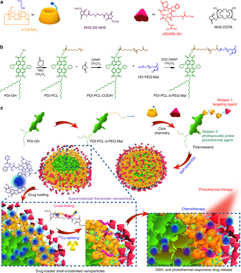
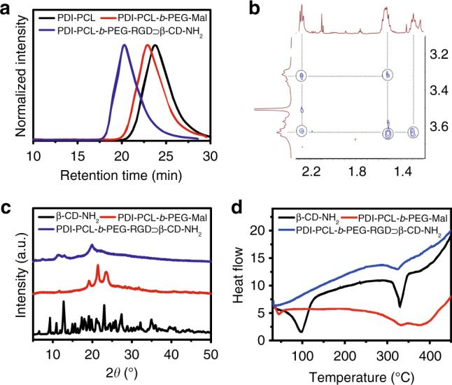
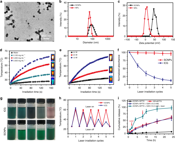
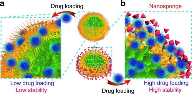
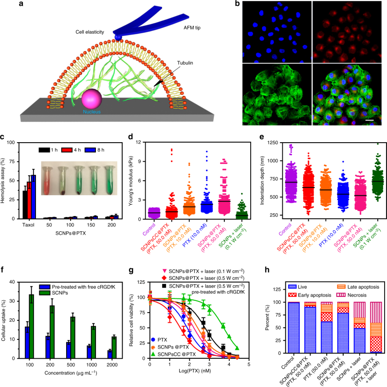
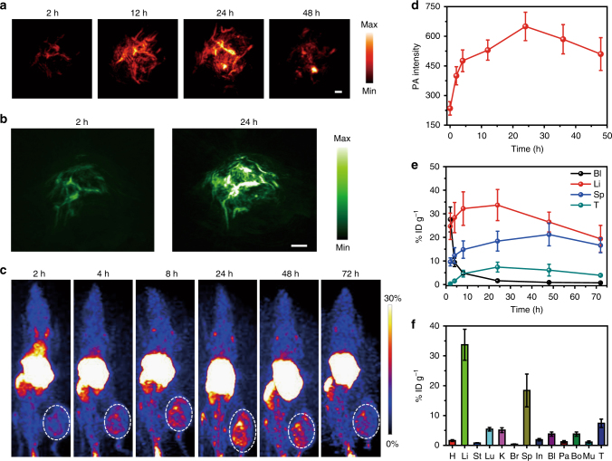
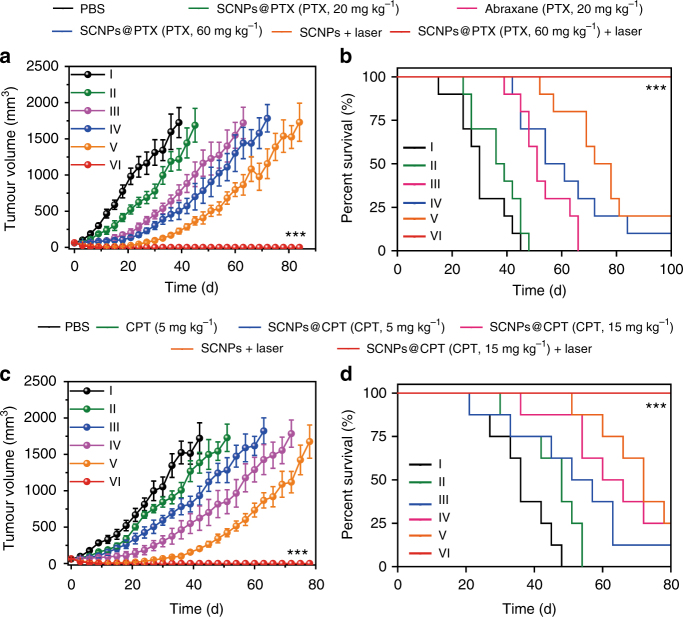
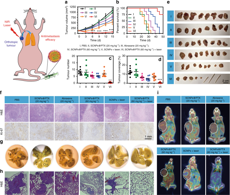
The development of smart theranostic systems with favourable biocompatibility, high loading efficiency, excellent circulation stability, potent anti-tumour activity, and multimodal diagnostic functionalities is of importance for future clinical application. The premature burst release and poor degradation kinetics indicative of polymer-based nanomedicines remain the major obstacles for clinical translation. Herein we prepare theranostic shell-crosslinked nanoparticles (SCNPs) using a β-cyclodextrin-based polyrotaxane (PDI-PCL-b-PEG-RGD⊃β-CD-NH2) to avoid premature drug leakage and achieve precisely controllable release, enhancing the maximum tolerated dose of the supramolecular nanomedicines. cRGDfK and perylene diimide are chosen as the stoppers of PDI-PCL-b-PEG-RGD⊃β-CD-NH2, endowing the resultant SCNPs with excellent integrin targeting ability, photothermal effect, and photoacoustic capability. In vivo anti-tumour studies demonstrate that drug-loaded SCNPs completely eliminate the subcutaneous tumours without recurrence after a single-dose injection combining chemotherapy and photothermal therapy. These supramolecular nanomedicines also exhibit excellent anti-tumour performance against orthotopic breast cancer and prevent lung metastasis with negligible systemic toxicity.


Conflict of interest statement

The authors declare no competing financial interests.

Figures

Fig. 1
Fig. 1
Synthesis and fabrication schematics of SCNPs for supramolecular theranostics. a Chemical structures and cartoon representations of the building blocks (β-CD-NH2, NHS-SS-NHS, cRGDfK-SH, and NHS-DOTA). b Synthetic route to the polyrotaxane (PDI-PCL-b-PEG-RGD⊃β-CD-NH2). c Schematic illustrations of the preparation of drug-loaded SCNPs and dual-responsive drug release
Fig. 2
Fig. 2
Characterisation of polyrotaxane. a GPC curves of PDI-PCL, PDI-PCL-b-PEG-Mal, and PDI-PCL-b-PEG-RGD⊃β-CD-NH2. b 2D NOESY NMR spectrum (500 MHz, 298 K) of PDI-PCL-b-PEG-RGD⊃β-CD-NH2. c XRD patterns of β-CD-NH2, PDI-PCL-b-PEG-Mal, and PDI-PCL-b-PEG-RGD⊃β-CD-NH2. d DSC thermograms of β-CD-NH2, PDI-PCL-b-PEG-Mal, and PDI-PCL-b-PEG-RGD⊃β-CD-NH2
Fig. 3
Fig. 3
Preparation of SCNPs and stimuli-responsive drug release. a TEM image of SCNPs. b DLS results and c zeta potential of the NPs formed from PDI-PCL-b-PEG-RGD⊃β-CD-NH2 and SCNPs. d The photothermal curves of pure water and SCNPs at different concentrations under 671 nm laser irradiation at a power density of 0.5 W cm−2. e The photothermal curves of SCNPs (0.250 mg mL−1) under 671 nm laser irradiation at different power densities. f The changes in absorbance intensity, g photographs, and h thermal curves of SCNPs and ICG after five cycles of irradiation, respectively. The test laser wavelengths of SCNPs and ICG were 671 and 780 nm, respectively, with a power density of 0.5 W cm−2. i Controlled release profiles of SCNPs@PTX and NPs@PTX under different conditions. Data are expressed as means ± s.e.m. (n = 3)
Fig. 4
Fig. 4
Schematic illustration of drug encapsulation. Schematic comparison of drug loading efficiency and stability between a NPs and b SCNPs formed from PDI-PCL-b-PEG-Mal and polyrotaxane, respectively
Fig. 5
Fig. 5
In vitro thermo-chemotherapy. a Schematic of AFM method used in the measurements of cell mechanical properties. b CLSM images of the HeLa cells cultured with Cy5.5-labelled SCNPs@PTX. Blue fluorescence shows nuclear staining with Hoechst 33342; red fluorescence shows the location of Cy5.5-labelled SCNPs@PTX; green fluorescence shows β-actin staining with FITC-phalloidin. Scale bar is 20 μm. c Haemolysis rates of PTX (50 μg mL−1) and SCNPs@PTX at various concentrations (50, 100, 150, and 200 μg PTX mL−1). Inset: the corresponding image of RBCs treated with PTX and SCNPs@PTX at various concentrations for 8 h. Histogram of d reduced Young’s modulus and e indentation depth for all data collected from six different groups. The irradiation time was 3 min. f Cellular uptake of SCNPs@PTX under various concentrations in the absence and presence of free cRGDfK (20 μM). g In vitro cytotoxicity of different formulations towards HeLa cells. h Flow-cytometric analysis of Annexin-V/PI staining of HeLa cells after different treatments. The laser density was 0.5 W cm−2, and the irradiation time was 3 min. Data are expressed as means ± s.e.m. (n = 5)
Fig. 6
Fig. 6
In vivo PA and PET imaging. a Representative PA maximum imaging projection (MIP) and b 3D images of tumour in a living mouse after systemic administration of SCNPs through i.v. injection. Scale bar is 2 mm. c Decay-corrected whole-body coronal PET images of HeLa tumour-bearing mice at 2, 4, 8, 24, 48, and 72 h after i.v. injection of 150 μCi of 64Cu SCNPs. d Quantification of PA intensities at 671 nm as a function of post-injection time of SCNPs (n = 3). e Time-activity curves quantified based on PET images (n = 3). f Biodistribution of the 64Cu SCNPs in mice bearing HeLa tumours at 24 h post-injection (n = 3). Data are expressed as means ± s.e.m
Fig. 7
Fig. 7
In vivo thermo-chemotherapy on xenograft tumours. a Tumour volume changes and b Kaplan–Meier survival curves of the mice bearing HeLa xenografts treated with different formulations after one injection (n = 10). c Tumour volume changes and d Kaplan–Meier survival curves of mice bearing A549 xenografts treated with different formulations after one injection (n = 8). The laser density was 0.5 W cm−2, and the irradiation time was 5 min. Data are expressed as means ± s.e.m., ***P < 0.001
Fig. 8
Fig. 8
Treatment of orthotopic breast cancer and inhibitory effects on lung metastasis. a Tumour volume changes and b Kaplan–Meier survival curves of the mice bearing orthotopic 4T1 breast tumours treated with different formulations after one injection (n = 8). c The numbers of tumour nodules present on the lung surface from each group. d Tumour coverage percentage in the lungs from each group. e Photo images of the orthotopic tumours harvested from the mice treated with different formulations. f H&E and Ki-67 staining of the tumour tissues from each group. g Representative images of the lungs excised from each group. The black circles denote the visually detected metastatic nodules in each lung tissue. h Histological examination of metastatic lesions in lung tissues from each group after H&E staining. i PET/CT images of the mice treated with different formulations at the 14th day post-injection. Arrows indicates possible metastatic tumours. The laser density was 0.5 W cm−2, and the irradiation time was 5 min. Data are expressed as means ± s.e.m., ***P < 0.001

References

    1. Torchilin VP. Multifunctional, stimuli-sensitive nanoparticulate systems for drug delivery. Nat. Rev. Drug Discov. 2014;13:813–827. doi: 10.1038/nrd4333. - DOI - PMC - PubMed
    1. LaVan DA, McGuire T, Langer R. Small-scale systems for in vivo drug delivery. Nat. Biotechnol. 2003;21:1184–1191. doi: 10.1038/nbt876. - DOI - PubMed
    1. Petros RA, DeSimone JM. Strategies in the design of nanoparticles for therapeutic applications. Nat. Rev. Drug Discov. 2010;9:615–627. doi: 10.1038/nrd2591. - DOI - PubMed
    1. Elsabahy M, Heo GS, Lim SM, Sun G, Wooley KL. Polymeric nanostructures for imaging and therapy. Chem. Rev. 2015;115:10967–11011. doi: 10.1021/acs.chemrev.5b00135. - DOI - PMC - PubMed
    1. Elsabahy M, Wooley KL. Data mining as a guide for the construction of cross-linked nanoparticles with low immunotoxicity via control of polymer chemistry and supramolecular assembly. Acc. Chem. Res. 2015;48:1620–1630. doi: 10.1021/acs.accounts.5b00066. - DOI - PMC - PubMed

Publication types

MeSH terms

LinkOut - more resources