. 2018 Feb 22;8(1):3508.
doi: 10.1038/s41598-018-21974-x.

BigR is a sulfide sensor that regulates a sulfur transferase/dioxygenase required for aerobic respiration of plant bacteria under sulfide stress

Affiliations

BigR is a sulfide sensor that regulates a sulfur transferase/dioxygenase required for aerobic respiration of plant bacteria under sulfide stress

Nayara Patricia Vieira de Lira et al. Sci Rep. .

Abstract

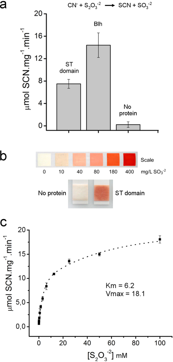
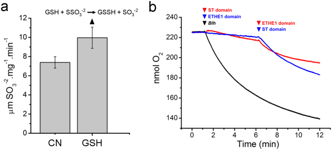
To cope with toxic levels of H2S, the plant pathogens Xylella fastidiosa and Agrobacterium tumefaciens employ the bigR operon to oxidize H2S into sulfite. The bigR operon is regulated by the transcriptional repressor BigR and it encodes a bifunctional sulfur transferase (ST) and sulfur dioxygenase (SDO) enzyme, Blh, required for H2S oxidation and bacterial growth under hypoxia. However, how Blh operates to enhance bacterial survival under hypoxia and how BigR is deactivated to derepress operon transcription is unknown. Here, we show that the ST and SDO activities of Blh are in vitro coupled and necessary to oxidize sulfide into sulfite, and that Blh is critical to maintain the oxygen flux during A. tumefaciens respiration when oxygen becomes limited to cells. We also show that H2S and polysulfides inactivate BigR leading to operon transcription. Moreover, we show that sulfite, which is produced by Blh in the ST and SDO reactions, is toxic to Citrus sinensis and that X. fastidiosa-infected plants accumulate sulfite and higher transcript levels of sulfite detoxification enzymes, suggesting that they are under sulfite stress. These results indicate that BigR acts as a sulfide sensor in the H2S oxidation mechanism that allows pathogens to colonize plant tissues where oxygen is a limiting factor.


Conflict of interest statement

The authors declare no competing interests.

Figures

Figure 1
Figure 1
The ETHE1 domain of Blh is an iron-containing sulfur dioxygenase. (a) SDO activity of the Blh ETHE1 domain (black line), relative to buffer with no protein (red line), according to the reaction scheme shown. Oxygen consumption, as a measure of the SDO activity of the Blh ETHE1 domain, occurs only after the addition of substrate GSSH to the reaction mixture (arrow). (b) Sulfite production at the terminus of the SDO reaction estimated by the sulfite test strip, according to the scale. (c) Average O2 concentration per mg of protein (ETHE1 domain) per min at 25 °C, relative to control (no protein in the reaction mixture). Values are the mean of three independent measurements and error bars indicate standard deviations. (d) SDO activity as function of the GSSH concentration, showing that the ETHE1 domain displays a Michaelis-Menten kinetics with an estimated apparent Km and Vmax values around 0.18 mM GSSH and 3.4 µmol O2 mg−1 min−1, respectively. (e) X-ray fluorescence analysis showing higher levels of iron in the ETHE1 domain sample, relative to buffer alone (no protein) or BSA, as negative control samples.
Figure 2
Figure 2
The Blh DUF442 domain is a sulfur transferase. (a) ST activity of the full-length Blh or its DUF442 domain alone (ST domain), relative to buffer with no protein. The ST activity, as a measure of iron-SCN formation, used thiosulfate as the sulfur donor and cyanide as the sulfur acceptor, according to the reaction scheme shown. Values are the mean of nine independent measurements and error bars indicate the standard deviation. (b) Sulfite production at the terminus of the ST reaction estimated by the sulfite test strip, according to the scale, indicating that both the full-length Blh and DUF442 domain catalyzed the transfer of the sulfane sulfur of thiosulfate to cyanide, generating SCN and sulfite as the reaction products. (c) ST activity of full-length Blh as function of the thiosulfate concentration, showing that Blh displays a Michaelis-Menten kinetics with an estimated apparent Km and Vmax values of ~6.2 mM thiosulfate and 18.1 µmol SCN mg−1 min−1, respectively.
Figure 3
Figure 3
The couple ST and SDO activities of Blh. (a) ST activity of the Blh ST domain as a measure of sulfite production when cyanide (CN) or GSH are used as sulfur acceptors and thiosulfate as the sulfur donor. Values are the mean of three independent measurements and error bars indicate the standard deviation. In addition to sulfite, the ST activity upon GSH is expected to generate GSSH, according to the reaction scheme shown. (b) O2 consumption as a measure of the SDO activity of the Blh ETHE1 domain showing that the O2 concentration in the medium is not significantly altered when the ST domain is added to the reaction mixture containing thiosulfate and GSH. Nevertheless, O2 consumption increases significantly soon after the addition of the ETHE1 domain to the reaction mixture (red line). Similar results are observed when the ETHE1 domain is added first to the reaction mixture (blue line). When the full-length Blh is used, a sharper decrease in the O2 levels is observed (black line), indicating that the ST and SDO activities of Blh are in vitro coupled.
Figure 4
Figure 4
Blh is required to maintain aerobic respiration under O2-limited conditions. O2 consumption (black lines) and O2 flux (red lines), as a measure of aerobic respiration, exhibited by the wild type (a) A. tumefaciens and corresponding blh (b) and bigR (c) cells in medium containing glucose. The blh cells showed a lower O2 flux (b) compared to the wild type cells (a) under O2-replete conditions (above 20 nmol mL−1), whereas the bigR cells (c) showed a higher O2 flux relative to the wild type cells. (d) O2 flux as a function of the O2 concentration showing that the differences in the O2 flux between the blh and bigR mutants, relative to the wild type cells, increased significantly as the O2 concentration in the medium approaches zero. The O2 flux of blh cells at 0.2 nmol mL−1 O2 was approximately 10 × lower than that of the wild type cells, but nearly the same of that of the wild type cells at 5.0 nmol mL−1 O2. Conversely, the bigR cells maintained a high O2 flux even at 0.2 nmol mL−1 O2, compared to the wild type and blh cells.
Figure 5
Figure 5
Xylella-infected plants are under sulfite stress. (a) Leaf discs of Pineapple sweet orange incubated with increased amounts of sulfite or sulfate, showing that sulfite, but not sulfate, induces chlorosis in citrus. (b) Example of a symptomatic CVC leaf of Pineapple plants showing typical CVC lesions surrounded by chlorotic halos (left panel), compared with an asymptomatic leaf (right panel). (c) Sulfite levels measured in symptomatic (S) and asymptomatic (A) CVC leaves showing higher sulfite levels in symptomatic and asymptomatic leaves relative to non-infected control (C) plants. Values are the means of six independent measurements and error bars indicate the standard deviation, whereas asterisks indicate statistically significant difference between the means. (d) Quantitative PCR analysis showing high bacterial titles in both symptomatic and asymptomatic leaves of CVC plants. No PCR amplification is detected in non-infected plants. (e) Quantitative PCR analysis showing the relative mRNA levels of SiR and SO between symptomatic and asymptomatic CVC leaves, compared to non-infected control plants. Values in ‘d’ and ‘e’ are the means of five independent biological replicates and asterisks indicate statistically significant difference between the means.
Figure 6
Figure 6
H2S and polysulfide oxidizes BigR and activates the bigR operon. (a) Gel-shift assay stained with ethidium bromide showing formation of the BigR-DNA complex (arrow) in the control mix reaction (C), or in the presence of GSH. The BigR-DNA complex is not only abolished in the presence of GSSH, but also in the presence of acetonic sulfur (S), used in combination with GSH to generate GSSH. FP designates the free probe. (b) Detection of HS and polysulfides in the commercial source of elemental sulfur. Acetone adjusted to pH 12 with potassium hydroxide was used as a control solution (C). Addition of sulfur (S) caused the solution to turn yellow indicating the presence of polysulfides in the sample. Addition of bismuth citrate (Bi) to the acetone solution formed a white precipitate which turned into an orange-brownish color with sulfur addition (Bi + S), indicating the presence of HS in the sample. (c) Absorbance curve of the ammonium sulfide solution (60 mM) in the presence and absence of sulfur showing peaks at 300 and 375 nm characteristic of polysulfides. (d) Gel-shift assay showing that the BigR protein treated with H2S gas no longer binds DNA. (e) Fluorescence images of A. tumefaciens cells carrying the GFP reporter plasmid for the bigR operon growing in the presence and absence of the polysulfide solution at 0.2%, 2.0% or 10% v/v. Cells exposed to the polysulfide solution show increased GFP fluorescence in a dose-dependent manner, indicating that HS and/or polysulfides are responsible for the operon activation.

Similar articles

Cited by

References

    1. Paul BD, Snyder SH. H2S signaling through protein sulfhydration and beyond. Nat. Rev. Mol. Cell Biol. 2012;13:499–407. doi: 10.1038/nrm3391. - DOI - PubMed
    1. Stein A, Bailey SM. Redox biology of hydrogen sulfide: implications for physiology, pathophysiology, and pharmacology. Redox Biol. 2013;1:32–39. doi: 10.1016/j.redox.2012.11.006. - DOI - PMC - PubMed
    1. Li ZG, Min X, Zhou ZH. Hydrogen sulfide: a signal molecule in plant cross-adaptation. Front. Plant Sci. 2016;7:1621. - PMC - PubMed
    1. Clarke PH. Hydrogen sulphide production by bacteria. J. Gen. Microbiol. 1953;8:397–407. doi: 10.1099/00221287-8-3-397. - DOI - PubMed
    1. Barrett EL, Clark MA. Tetrathionate reduction and production of hydrogen sulfide from thiosulfate. Microbiol. Rev. 1987;51:192–205. - PMC - PubMed

Publication types

LinkOut - more resources