. 2018 Feb 23;50(2):e449.
doi: 10.1038/emm.2017.281.

Inhibition of TNFα-interacting protein α (Tipα)-associated gastric carcinogenesis by BTG2/TIS21 via downregulating cytoplasmic nucleolin expression

Affiliations

Inhibition of TNFα-interacting protein α (Tipα)-associated gastric carcinogenesis by BTG2/TIS21 via downregulating cytoplasmic nucleolin expression

Preethi Devanand et al. Exp Mol Med. .

Abstract

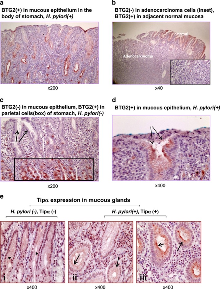
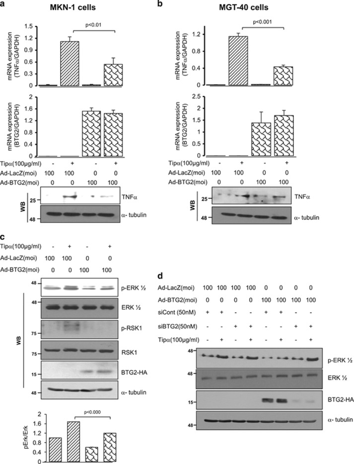
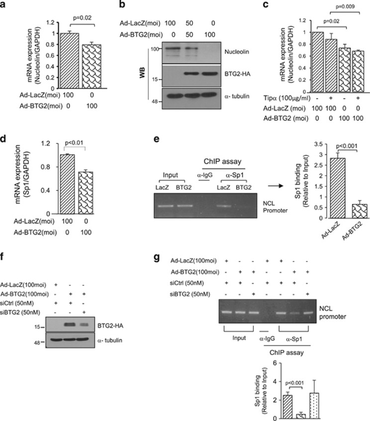
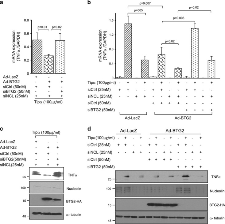
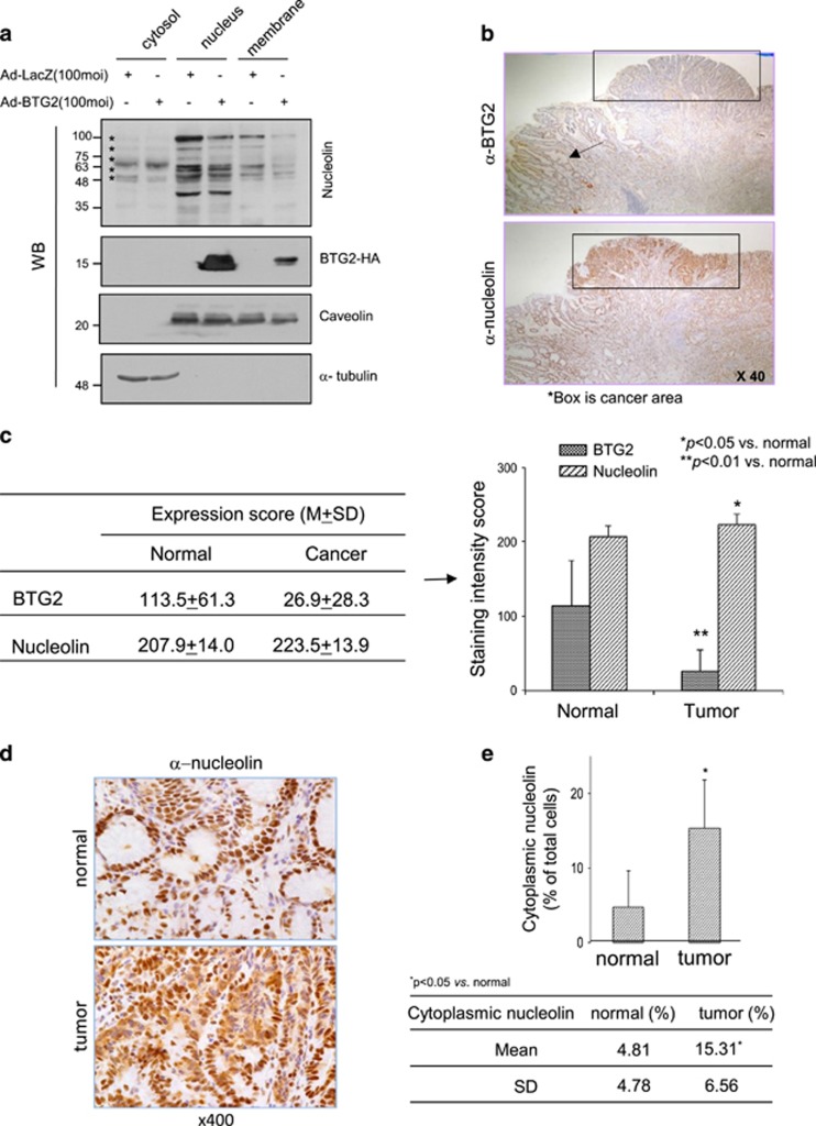
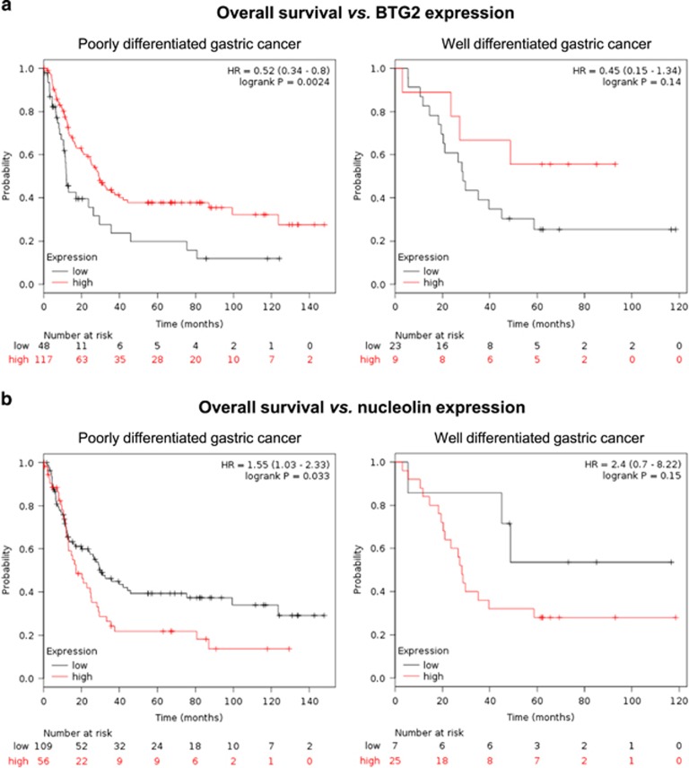
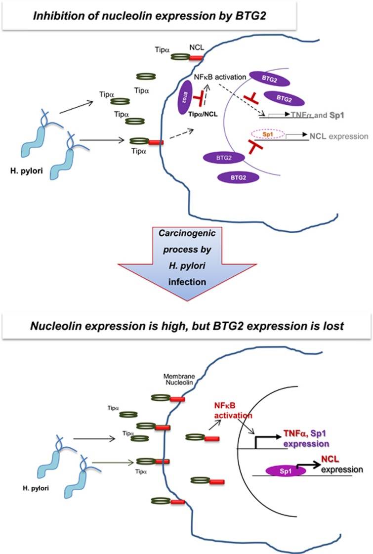
To understand the regulation of Helicobacter pylori (H. pylori)-associated gastric carcinogenesis, we examined the effect of B-cell translocation gene 2 (BTG2) expression on the biological activity of Tipα, an oncoprotein secreted from H. pylori. BTG2, the human ortholog of mouse TIS21 (BTG2/TIS21), has been reported to be a primary response gene that is transiently expressed in response to various stimulations. Here, we report that BTG2 is constitutively expressed in the mucous epithelium and parietal cells of the gastric gland in the stomach. Expression was increased in the mucous epithelium following H. pylori infection in contrast to its loss in human gastric adenocarcinoma. Indeed, adenoviral transduction of BTG2/TIS21 significantly inhibited Tipα activity in MKN-1 and MGT-40, human and mouse gastric cancer cells, respectively, thereby downregulating tumor necrosis factor-α (TNFα) expression and Erk1/2 phosphorylation by reducing expression of nucleolin, a Tipα receptor. Chromatin immunoprecipitation proved that BTG2/TIS21 inhibited Sp1 expression and its binding to the promoter of the nucleolin gene. In addition, BTG2/TIS21 expression significantly reduced membrane-localized nucleolin expression in cancer cells, and the loss of BTG2/TIS21 expression induced cytoplasmic nucleolin availability in gastric cancer tissues, as evidenced by immunoblotting and immunohistochemistry. Higher expression of BTG2 and lower expression of nucleolin were accompanied with better overall survival of poorly differentiated gastric cancer patients. This is the first report showing that BTG2/TIS21 inhibits nucleolin expression via Sp1 binding, which might be associated with the inhibition of H. pylori-induced carcinogenesis. We suggest that BTG2/TIS21 is a potential inhibitor of nucleolin in the cytoplasm, leading to inhibition of carcinogenesis after H. pylori infection.


Conflict of interest statement

The authors declare no conflict of interest.

Figures

Figure 1
Figure 1
BTG2/TIS21 expression is increased in normal mucous epithelium with H. pylori infection, but absent in stomach cancer cells. Immunohistochemistry (IHC) findings of BTG2/TIS21 expression on the serial sections of normal mucosa and cancer tissues of the human stomach infected with H. pylori. (a) BTG2 expression was increased in the neck portion of gastric glands in the body of the stomach with H. pylori infection, × 200. (b) BTG2 expression was lost in adenocarcinoma despite the mild expression in normal mucous epithelium, × 40. Inset is the high-power view revealing no BTG2 expression in carcinoma cells. (c) Absence of BTG2 expression in mucous epithelium (arrows) compared with strong positive expression in the parietal cells (rectangle) of the human stomach without H. pylori infection, × 200. (d) Induction of BTG2 expression in the surface of mucous glands infected with many H. pylori (arrows), × 400. (e) To confirm the secretion of Tipα and H. pylori infection, serial sections of paraffin blocks were stained. Note mild (panel ii) to strong (panel iii) expression of Tipα and H. pylori in the mucous glands of the stomach with H. pylori (arrows) infection but no Tipα expression (panel i) in the mucous glands of the stomach without H. pylori infection (arrow heads), × 400.
Figure 2
Figure 2
Tipα activity is significantly reduced by BTG2 expression in human gastric cancer cells. To evaluate the effect of BTG2 expression on Tipα activity, Tipα-induced TNFα expression and ERK1/2 activation were examined in the human and mouse gastric cancer cells MKN-1 (a) and MGT-40 (b), respectively, after transduction of the cells with 100 MOI of adenovirus carrying the LacZ or BTG2 gene for 5 h. The cells were maintained until 48 h before treatment with either Tipα (100 μg ml−1) or vehicle for 1 h. Cellular RNAs were isolated to examine the regulation of TNFα expression by RT-qPCR. GAPDH expression was used as an internal control. Immunoblot analysis was also performed using the above samples, TNFα protein expression was analyzed, and α-tubulin served as a loading control. Tipα-induced TNFα expression was significantly reduced in the BTG2 expresser compared with that in the LacZ control. (c) Immunoblot (IB) analysis and quantitation. Tipα-induced ERK1/2 activation was significantly reduced in MKN-1 cells with BTG2 expression compared with that in the LacZ control. pRSK1, a downstream target of pERK1/2, was also reduced. α-Tubulin serves as a loading control (upper panel). Densitometric analysis was performed using ImageJ software (Lower panel); regulation of Tipα-induced pERK1/2 in Ad-LacZ was significantly reduced in the Ad-BTG2-infected cells. (d) Regulation of Tipα-induced ERK1/2 activation by the knockdown of BTG2 expression. To confirm the effect of BTG2 expression on the downregulation of Tipα-induced ERK1/2 activation, MKN-1 cells were transfected with siBTG2 before transduction with Ad-BTG2, and then the cells were subjected to IB analysis. Note the recovery of the BTG2-inhibited pERK1/2 level by siBTG2 transfection.
Figure 3
Figure 3
Expression of nucleolin, a Tipα receptor, is reduced by BTG2 expression via the inhibition of Sp1 binding to the nucleolin promoter. To investigate whether BTG2 regulates the expression of the Tipα receptor, nucleolin (NCL) mRNA was isolated from MKN-1 cells infected with either Ad-LacZ or Ad-BTG2 before the following analyses: (a) RT-qPCR, in which BTG2 reduced NCL mRNA expression in MKN-1 cells compared with that in LacZ-transduced cells. GAPDH was used as an internal control. (b) IB analysis, in which BTG2 significantly reduced NCL protein expression. α-Tubulin served as a loading control. (c) RT-qPCR analysis, in which NCL expression was lower in the BTG2 expresser than in the LacZ expresser independent of Tipα treatment. (d) RT-qPCR analysis revealed the inhibition of Sp1 expression in MKN-1 cells with BTG2 expression. GAPDH was internal control. (e) Chromatin immunoprecipitation (ChIP) assay. Sp1 binding to the NCL promoter was reduced in the BTG2-transduced MKN-1 cells compared with that in the LacZ-transduced cells. Immunoprecipitation (IP) with normal IgG was employed as the IP control (Left panel). ImageJ analysis was used to analyze Sp1 binding to the NCL promoter in the LacZ- and BTG2-transduced cells. BTG2 transduction significantly downregulated Sp1 binding to the NCL promoter (right panel). (f) IB analysis revealed the knockdown of BTG2 expression by transfection of MKN-1 cells with siBTG2. siControl RNAs were obtained from the scrambled sequences. (g) ChIP assay confirmed the activity of the BTG2 gene in Sp1 binding to the NCL promoter (upper panel). ImageJ analysis was applied, and we found that Sp1 binding was significantly reduced in the BTG2 expresser but was recovered by transfection with siBTG2 (lower panel).
Figure 4
Figure 4
Tipα-induced TNFα expression is inversely regulated by nucleolin and BTG2. To confirm the effect of BTG2 expression on Tipα-induced TNFα expression, endogenous NCL expression was excluded by transfection of MKN-1 cells with siNCL, and then adenoviral transduction was performed. RT-qPCR analysis (a) Tipα-induced TNFα expression was significantly reduced in the BTG2 expresser, but it was recovered by the knockdown of BTG2 with siBTG2 transfection. Expression of GAPDH served as the internal control. (c) IB analysis clearly revealed the regulation of TNFα expression depending on BTG2 expression. Knockdown of nucleolin and BTG2-HA was also revealed. α-tubulin served as an internal control. To explore the effect of NCL on the regulation of Tipα-induced TNFα expression, MKN-1 cells transfected with either siNCL or siBTG2 were transduced with either Ad-LacZ or Ad-BTG2, and then Tipα was treated 1 h before RNA extraction. (b) Note the significant inhibition of Tipα-induced TNFα expression by the knockdown of NCL expression in both the LacZ and BTG2 expressers, indicating the induction of TNFα expression by NCL expression. (d) Immunoblot analysis was performed to analyze the protein expressions of TNFα, nucleolin and BTG2-HA in the present experiment. α-Tubulin served as an internal control. The data strongly suggest that BTG2 and NCL inversely regulate Tipα activity in gastric cancer cells.
Figure 5
Figure 5
Gastric cancer expresses nucleolin in the cytoplasm but has lost BTG2 expression. (a) IB analysis. MKN-1 cells transduced with either Ad-LacZ or Ad-BTG2 were fractionated into cytosolic, nuclear and membranous parts and were subjected to IB analysis. NCL expression was downregulated in the membrane fraction of the BTG2 expresser. *indicates NCL proteins detected by IB analysis. α-Tubulin served as a loading control for the cytosol, and caveolin served as the control for the nuclear and membranous fractions. The data represent three independent trials. (b) IHC of human gastric cancer tissues with anti-BTG2 (upper) and anti-NCL (lower) antibodies. BTG2 expression was absent in gastric carcinoma (rectangle) but was still present in normal tissue (arrow). By contrast, NCL expression was strong in the carcinoma (rectangle) compared with that in normal tissue. (c) The extent of BTG2 and NCL expression was scored after staining tumor tissues with anti-BTG2 and anti-NCL antibodies, and then the staining intensity was scored from 0+ to 3+ as shown in Supplementary Figure 1c. BTG2 expression was four times higher in normal than in tumor tissues (p<0.01), whereas NCL expression was higher in tumor than in normal tissues (p<0.05), indicating the loss of BTG2 expression as opposed to the gain of NCL expression in cancer tissues compared with control tissues. (d) IHC with anti-NCL antibody; cytoplasmic expression of NCL was noted in cancer cells but not in normal mucous epithelium. The upper panel shows NCL expression in the nuclei of the gastric gland epithelium in the normal stomach. The lower panel reveals strong expression of NCL in both the cytoplasm and nuclei of cancer cells. (e) NCL expression in the cytoplasm and nuclei was counted under a microscope and was expressed as % of the total cells, *p< 0.05 vs normal. Note the significant increase in the cells with NCL expression out of the nuclei. Supplementary Figure 1d shows representative cells with cytoplasmic NCL expression used for the staining score.
Figure 6
Figure 6
Overall survival of gastric cancer is reciprocally regulated by the expression of BTG2/TIS21 and nucleolin. To explore the clinical significance and correlation between the survival of gastric cancer and expression of BTG2 and NCL, we examined the public databases concerning human gastric cancer. The red line indicates the high expresser, and the black one indicates the low expresser. Kaplan–Meier analysis showing the overall survival vs BTG2 and NCL expressions in gastric cancer patients. (a) Note the better survival rate in the poorly differentiated gastric cancer patients with BTG2 high expression than in those with BTG2 low expression (p=0.0024), whereas it was statistically insignificant in the well-differentiated gastric cancers. (b) By contrast, the NCL high expresser exhibited a lower survival rate in the poorly differentiated gastric cancers compared with the low expressers (p=0.033). The correlation was also statistically insignificant in the well-differentiated gastric cancers. The numbers indicate the surviving patients at each time point.
Figure 7
Figure 7
BTG2/TIS21 downregulates Helicobacter pylori-associated gastric carcinogenesis by inhibiting Tipα activity via downregulating nucleolin expression by Sp1. Schema depicting a potential role of BTG2/TIS21 in gastric carcinogenesis. BTG2/TIS21 secreted from gastric gland epithelium inhibits Tipα-induced NFκB activation and its downstream activities; thus TNFα and Sp1 expression is inhibited. Consequently, NCL expression is downregulated due to the inhibition of Sp1 expression and its binding to the NCL promoter by BTG2/TIS21. The carcinogenic process after H. pylori infection induces BTG2/TIS21 expression in the gastric gland epithelium; however, the cells with BTG2 expression face growth arrest as opposed to the selective growth advantage in adenocarcinoma without BTG2 expression. The cells with lost BTG2 expression fail to block Tipα-induced TNFα and Sp1 transcription; thus, Sp1 can enhance NCL transcription. NCL transport to the cytoplasmic fraction can act as the receptor of Tipα secreted by H. Pylori in the stomach.

References

    1. Ferlay J, Soerjomataram I, Dikshit R, Eser S, Mathers C, Rebelo M et al. Cancer incidence and mortality worldwide: sources, methods and major patterns in GLOBOCAN 2012. Int J Cancer 2015; 136: E359–E386. - PubMed
    1. Shin A, Kim J, Park S. Gastric cancer epidemiology in Korea. J Gastric Cancer 2011; 11: 135–140. - PMC - PubMed
    1. Hill AB. The environment and disease: association or causation? Proc R Soc Med 1965; 58: 295–300. - PMC - PubMed
    1. Wroblewski LE, Peek RM Jr, Wilson KT. Helicobacter pylori and gastric cancer: factors that modulate disease risk. Clin Microbiol Rev 2010; 23: 713–739. - PMC - PubMed
    1. Tummuru MK, Cover TL, Blaser MJ. Cloning and expression of a high-molecular-mass major antigen of Helicobacter pylori: evidence of linkage to cytotoxin production. Infect Immun 1993; 61: 1799–1809. - PMC - PubMed

Publication types

MeSH terms