. 2018 May;32(5):1135-1146.
doi: 10.1038/s41375-017-0005-9. Epub 2018 Feb 2.

Pharmacologic inhibition of STAT5 in acute myeloid leukemia

Affiliations

Pharmacologic inhibition of STAT5 in acute myeloid leukemia

Bettina Wingelhofer et al. Leukemia. 2018 May.

Abstract

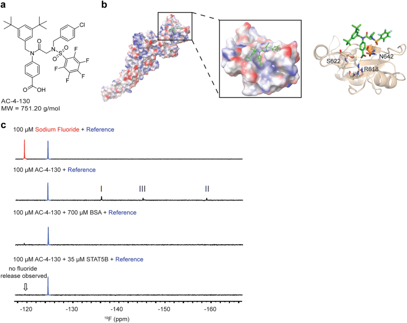
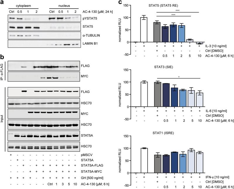
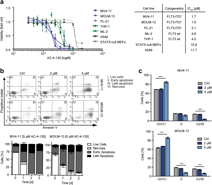
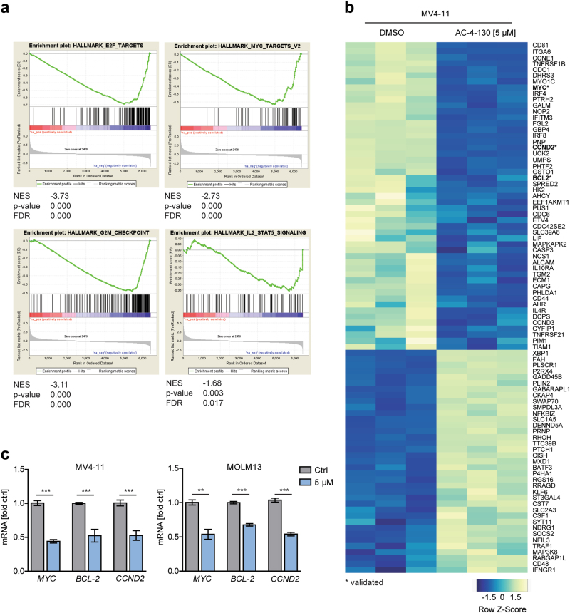
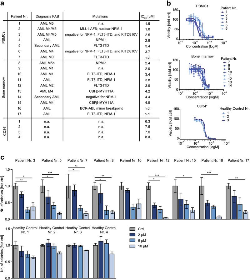
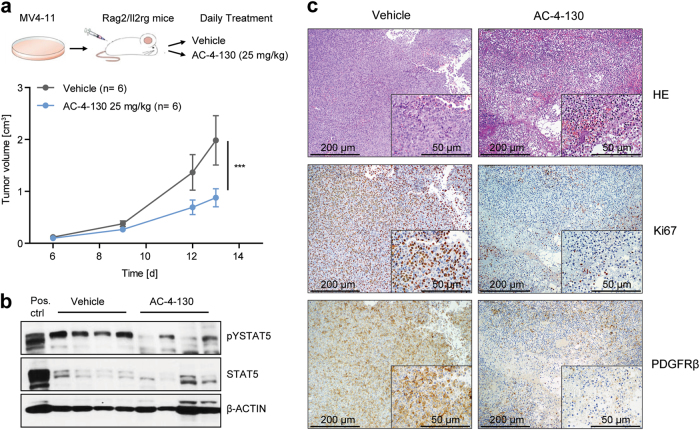
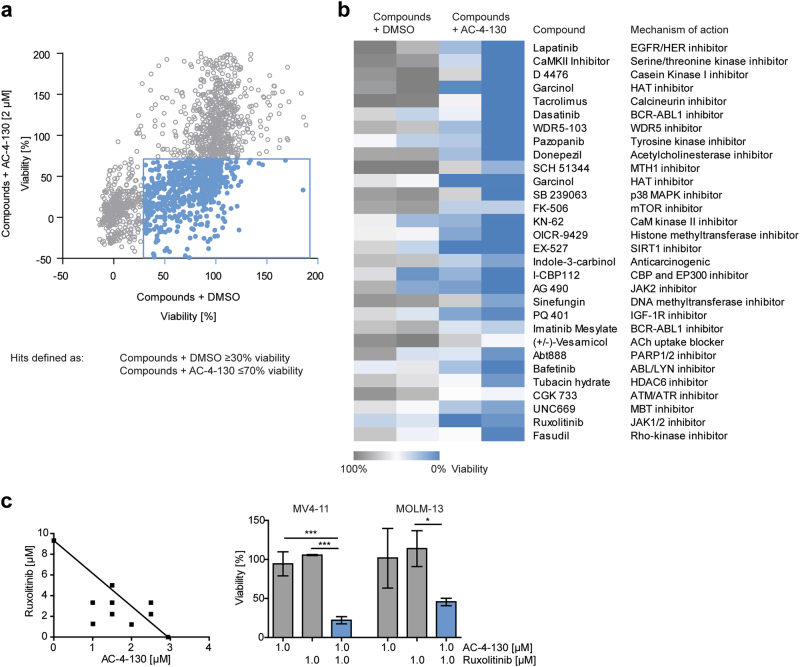
The transcription factor STAT5 is an essential downstream mediator of many tyrosine kinases (TKs), particularly in hematopoietic cancers. STAT5 is activated by FLT3-ITD, which is a constitutively active TK driving the pathogenesis of acute myeloid leukemia (AML). Since STAT5 is a critical mediator of diverse malignant properties of AML cells, direct targeting of STAT5 is of significant clinical value. Here, we describe the development and preclinical evaluation of a novel, potent STAT5 SH2 domain inhibitor, AC-4-130, which can efficiently block pathological levels of STAT5 activity in AML. AC-4-130 directly binds to STAT5 and disrupts STAT5 activation, dimerization, nuclear translocation, and STAT5-dependent gene transcription. Notably, AC-4-130 substantially impaired the proliferation and clonogenic growth of human AML cell lines and primary FLT3-ITD+ AML patient cells in vitro and in vivo. Furthermore, AC-4-130 synergistically increased the cytotoxicity of the JAK1/2 inhibitor Ruxolitinib and the p300/pCAF inhibitor Garcinol. Overall, the synergistic effects of AC-4-130 with TK inhibitors (TKIs) as well as emerging treatment strategies provide new therapeutic opportunities for leukemia and potentially other cancers.


Conflict of interest statement

The authors declare that they have no conflict of interest.

Figures

Fig. 1
Fig. 1
In vitro characterization of AC-4–130. a Chemical structure of AC-4–130. b Schematic representation of the STAT5B domain structure and binding mode of AC-4–130. c 1D 19F NMR studies of AC-4–130 with STAT5B
Fig. 2
Fig. 2
AC-4–130 inhibits STAT5 dimerization and target gene expression. a Subcellular fractions of Ba/F3 FLT3-ITD+ cells immunoblotted for pY-STAT5 and total STAT5. α-TUBULIN and LAMIN B1 were used as loading controls for cytoplasmic and nuclear fractions, respectively. Blots represent 2 independent experiments. Uncropped version of the Western blot is shown in Supplementary Fig. 8. b STAT5A-MYC and STAT5A-FLAG were co-transfected into HEK293T cells, co-immunoprecipitated with anti-FLAG and blotted with anti-FLAG and anti-MYC. Whole cell lysates were immunoblotted for MYC- or FLAG-tag, STAT5A, and HSC70 to show input. Results represent two independent experiments. Uncropped version of the Western blot is shown in Supplementary Fig. 8. c Ba/F3 cells were electroporated with Luciferase (Firefly) reporter plasmid for STAT5, and HT-29 cells were transfected with reporter plasmids for STAT1 or STAT3 in addition to pRL-TK (Renilla luciferase). Cells were starved, pretreated with AC-4–130 or DMSO (Ctrl) for 6 h and stimulation with appropriate cytokine. Relative luciferase activity was determined using the Dual-Luciferase Reporter Assay
Fig. 3
Fig. 3
FLT3-ITD+ cells are most susceptible to AC-4–130. a Viability assay for hematopoietic or control cell lines with AC-4–130 or DMSO (Ctrl) for 72 h. IC50 values (µM) were determined using GraphPad Prism 5 software (GraphPad Software, Inc.). b MV4–11 and MOLM-13 cells were treated with AC-4–130 or DMSO (Ctrl) in a dose-dependent manner for 72 h or with 5 µM AC-4–130 in a time-dependent manner. Apoptotic cells were detected by AnnexinV/PI staining. Representative dot plots are shown. c Cell cycle distribution was determined after 72 h using PI staining
Fig. 4
Fig. 4
RNA-seq analysis shows downregulation of the IL-2 STAT5 pathway. a GSEA of differentially expressed genes (p-value ≤ 0.01) in MV4–11 cells treated with AC-4–130 (5 µM) or DMSO (Ctrl) for 24 h. b Heatmap of differentially expressed genes in MV4–11 cells enriched in the IL-2 STAT5 hallmark pathway. c MV4–11 and MOLM-13 cells were treated with AC-4–130 or DMSO (Ctrl) for 24 h. mRNA expression of STAT5 target genes was analyzed by RT-qPCR. Data were normalized to GAPDH
Fig. 5
Fig. 5
AC-4–130 reduces clonogenicity of primary AML patient cancer stem cells. a Characteristics of human AML patients. b Viability assay of human AML patient samples and healthy CD34+ cells treated with AC-4–130 or DMSO (Ctrl) for 48 h. c AML samples and CD34+ cells were embedded in methylcellulose in the presence of AC-4–130 or DMSO (Ctrl). Colonies were counted 10 days after seeding
Fig. 6
Fig. 6
AC-4–130 decreases tumor formation and leukemogenesis in vivo. a Tumor volume of MV4–11 cells subcutaneously injected into both flanks of Rag2−/−γc−/− recipients, treated daily with vehicle or AC-4–130 (25 mg/kg). b Immunoblot showing pY-STAT5 and STAT5 levels after treatment. β-ACTIN was used as loading control. Uncropped version of the Western blot is shown in Supplementary Fig. 9. c H&E staining, Ki67, and PDGFRβ immunohistochemical staining of subcutaneously grown tumors
Fig. 7
Fig. 7
A chemical screen reveals compounds acting synergistically with AC-4–130. a MOLM-13 and MV4–11 cells were treated with a library of FDA-approved and experimental drugs (10–50 µM) alone or in combination with AC-4–130 (2 µM) for 72 h and cell viability was assessed. b Heatmap of hits defined as compounds giving a viability difference of 50% compared to DMSO controls. c Cell viability of MV4–11 and MOLM-13 cells treated with single drugs or combinations for 24 h. Synergy was assessed using Isobolograms

References

    1. Wang Z, Bunting KD. STAT5 in hematopoietic stem cell biology and transplantation. Jak-Stat. 2013;2:e27159. doi: 10.4161/jkst.27159. - DOI - PMC - PubMed
    1. Vainchenker W, Constantinescu SN. JAK/STAT signaling in hematological malignancies. Oncogene. 2013;32:2601–13. doi: 10.1038/onc.2012.347. - DOI - PubMed
    1. Hoelbl A, Schuster C, Kovacic B, Zhu B, Wickre M, Hoelzl MA, et al. Stat5 is indispensable for the maintenance of bcr/abl-positive leukaemia. EMBO Mol Med. 2010;2:98–110. doi: 10.1002/emmm.201000062. - DOI - PMC - PubMed
    1. Walz C, Ahmed W, Lazarides K, Betancur M, Patel N, Hennighausen L, et al. Essential role for Stat5a/b in myeloproliferative neoplasms induced by BCR-ABL1 and JAK2(V617F) in mice. Blood. 2012;119:3550–60. doi: 10.1182/blood-2011-12-397554. - DOI - PMC - PubMed
    1. Yan D, Hutchison RE, Mohi G. Critical requirement for Stat5 in a mouse model of polycythemia vera. Blood. 2012;119:3539–49. doi: 10.1182/blood-2011-03-345215. - DOI - PMC - PubMed

Publication types