

Altered Connectivity of the Anterior Cingulate and the Posterior Superior Temporal Gyrus in a Longitudinal Study of Later-life Depression
- PMID: 29472854
- PMCID: PMC5809471
- DOI: 10.3389/fnagi.2018.00031
Altered Connectivity of the Anterior Cingulate and the Posterior Superior Temporal Gyrus in a Longitudinal Study of Later-life Depression
Abstract
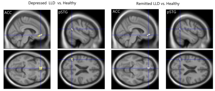
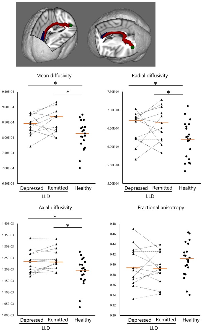
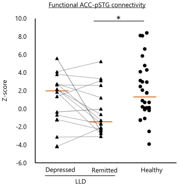
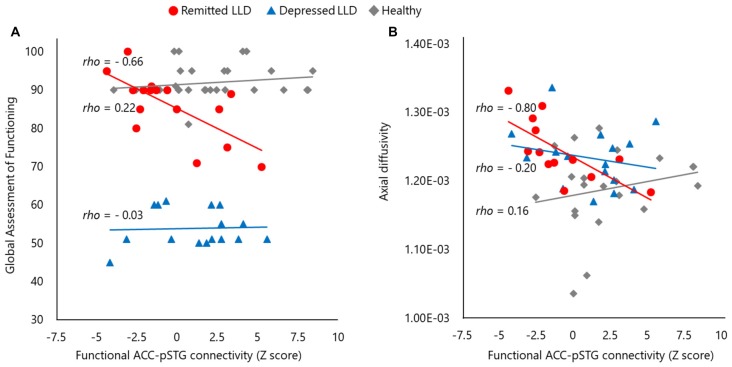
Patients with later-life depression (LLD) show abnormal gray matter (GM) volume, white matter (WM) integrity and functional connectivity in the anterior cingulate cortex (ACC) and posterior superior temporal gyrus (pSTG), but it remains unclear whether these abnormalities persist over time. We examined whether structural and functional abnormalities in these two regions are present within the same subjects during depressed vs. remitted phases. Sixteen patients with LLD and 30 healthy subjects were studied over a period of 1.5 years. Brain images obtained with a 3-Tesla magnetic resonance imaging (MRI) system were analyzed by voxel-based morphometry of the GM volume, and diffusion tensor imaging (DTI) and resting-state functional MRI were used to assess ACC-pSTG connectivity. Patients with LLD in the depressed and remitted phases showed significantly smaller GM volume in the left ACC and left pSTG than healthy subjects. Both patients with LLD in the depressed and remitted phases had significantly higher diffusivities in the WM tract of the left ACC-pSTG than healthy subjects. Remitted patients with LLD showed lower functional ACC-pSTG connectivity compared to healthy subjects. No difference was found in the two regions between depressed and remitted patients in GM volume, structural or functional connectivity. Functional ACC-pSTG connectivity was positively correlated with lower global function during remission. Our preliminary data show that structural and functional abnormalities of the ACC and pSTG occur during LLD remission. Our findings tentatively reveal the brain pathophysiology involved in LLD and may aid in developing neuroanatomical biomarkers for this condition.
Keywords: cingulate cortex; connectivity; gray matter volume; late-life depression; magnetic resonance imaging; resting state functional magnetic resonance imaging; superior temporal gyrus; white matter integrity.
Figures
Similar articles
-
Possible involvement of rumination in gray matter abnormalities in persistent symptoms of major depression: an exploratory magnetic resonance imaging voxel-based morphometry study.J Affect Disord. 2014 Oct;168:229-35. doi: 10.1016/j.jad.2014.06.030. Epub 2014 Jun 25. J Affect Disord. 2014. PMID: 25064808
-
Left posterior superior temporal gyrus and its structural connectivity in schizophrenia.Psychiatry Res Neuroimaging. 2025 Mar;347:111947. doi: 10.1016/j.pscychresns.2025.111947. Epub 2025 Jan 5. Psychiatry Res Neuroimaging. 2025. PMID: 39798501
-
Magnetic resonance imaging in late-life depression: multimodal examination of network disruption.Arch Gen Psychiatry. 2012 Jul;69(7):680-9. doi: 10.1001/archgenpsychiatry.2011.1862. Arch Gen Psychiatry. 2012. PMID: 22752234
-
Neural substrates for late-life depression: A selective review of structural neuroimaging studies.Prog Neuropsychopharmacol Biol Psychiatry. 2021 Jan 10;104:110010. doi: 10.1016/j.pnpbp.2020.110010. Epub 2020 Jun 13. Prog Neuropsychopharmacol Biol Psychiatry. 2021. PMID: 32544600 Review.
-
Evidence for Structural and Functional Alterations of Frontal-Executive and Corticolimbic Circuits in Late-Life Depression and Relationship to Mild Cognitive Impairment and Dementia: A Systematic Review.Front Neurosci. 2020 Apr 17;14:253. doi: 10.3389/fnins.2020.00253. eCollection 2020. Front Neurosci. 2020. PMID: 32362808 Free PMC article.
Cited by
-
Altered Intrinsic Brain Activity in Patients With Late-Life Depression: A Resting-State Functional MRI Study.Front Psychiatry. 2022 May 23;13:894646. doi: 10.3389/fpsyt.2022.894646. eCollection 2022. Front Psychiatry. 2022. PMID: 35677867 Free PMC article.
-
Common and distinct patterns of brain activity alterations during inhibitory control in depression and psychostimulant users: a comparative meta-analysis of task-based fMRI studies.Psychol Med. 2025 Jul 28;55:e218. doi: 10.1017/S0033291725101141. Psychol Med. 2025. PMID: 40717270 Free PMC article.
-
A comparative meta-analysis of structural magnetic resonance imaging studies and gene expression profiles revealing the similarities and differences between late life depression and mild cognitive impairment.Psychol Med. 2024 Nov 25;54(15):1-10. doi: 10.1017/S0033291724002563. Online ahead of print. Psychol Med. 2024. PMID: 39582389 Free PMC article.
-
Association of functional connectivity of the executive control network or default mode network with cognitive impairment in older adults with remitted major depressive disorder or mild cognitive impairment.Neuropsychopharmacology. 2023 Feb;48(3):468-477. doi: 10.1038/s41386-022-01308-2. Epub 2022 Apr 11. Neuropsychopharmacology. 2023. PMID: 35410366 Free PMC article.
-
Elevated homocysteine levels, white matter abnormalities and cognitive impairment in patients with late-life depression.Front Aging Neurosci. 2022 Jul 18;14:931560. doi: 10.3389/fnagi.2022.931560. eCollection 2022. Front Aging Neurosci. 2022. PMID: 35923546 Free PMC article.
References
LinkOut - more resources
Full Text Sources
Other Literature Sources