

Clinical and Magnetic Resonance Imaging Findings of Neurotoxocariasis
- PMID: 29472889
- PMCID: PMC5809457
- DOI: 10.3389/fneur.2018.00053
Clinical and Magnetic Resonance Imaging Findings of Neurotoxocariasis
Abstract
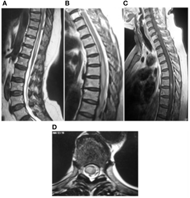
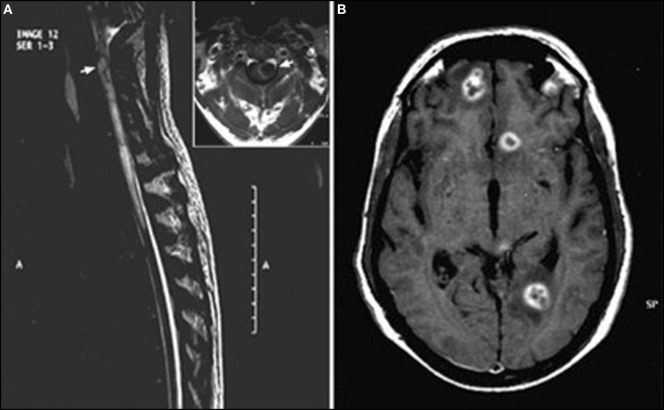
Human toxocariasis is one of the most prevalent helminthiases worldwide. Toxocara canis larvae can cross the blood-brain barrier leading to the neurotoxocariasis. The clinical presentation consists of a wide spectrum of neurological manifestations, but asymptomatic infection is probably common. Neurotoxocariasis is not a frequent diagnosis probably due to the non-specific nature of its symptoms as well as the lack of confirmatory diagnostic tests. Diagnosis of neurotoxocariasis is based on the presence of a high titer of anti-Toxocara antibody in the cerebrospinal fluid or in the serum, presence of eosinophilia in the serum or cerebrospinal fluid, and clinical and radiological improvement after anthelmintic therapy; however, universally accepted diagnostic criteria are lacking. Magnetic resonance imaging (MRI) findings include single or multiple, subcortical, cortical or white matter hyperintense lesions, best visualized on FLAIR and T2-weighted imaging, and usually isointense or hypointense on T1. These imaging findings are suggestive but not specific to neurotoxocariasis. Definitive diagnosis is made by histological confirmation, but it is rarely followed. This review provides an overview of the clinical manifestations, management options, and MRI findings of neurotoxocariasis.
Keywords: central nervous system infections; helminths; myelitis; neurotoxocariasis; toxocariasis.
Figures
References
Publication types
Grants and funding
LinkOut - more resources
Full Text Sources
Other Literature Sources