Review
. 2019 Feb 15;527(3):522-534.
doi: 10.1002/cne.24417. Epub 2018 Mar 9.

Binocular response modulation in the lateral geniculate nucleus

Affiliations
Review

Binocular response modulation in the lateral geniculate nucleus

Kacie Dougherty et al. J Comp Neurol. .

Abstract

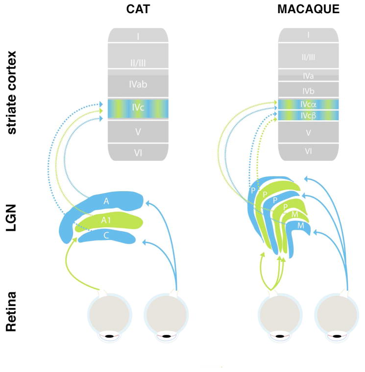
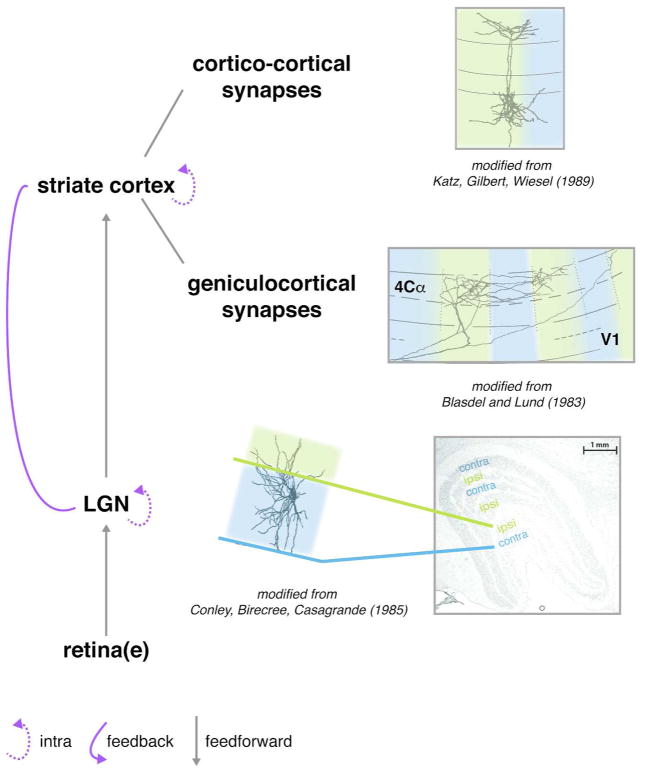
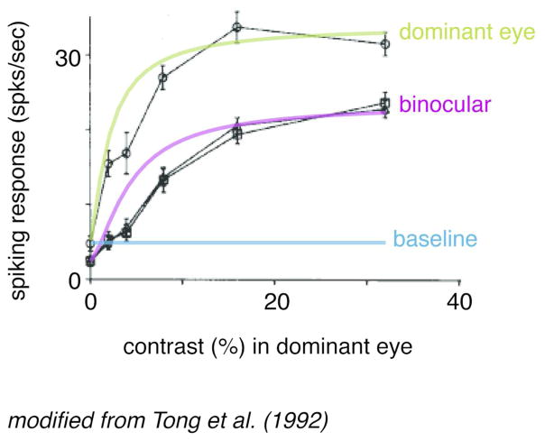
The dorsal lateral geniculate nucleus of the thalamus (LGN) receives the main outputs of both eyes and relays those signals to the visual cortex. Each retina projects to separate layers of the LGN so that each LGN neuron is innervated by a single eye. In line with this anatomical separation, visual responses of almost all of LGN neurons are driven by one eye only. Nonetheless, many LGN neurons are sensitive to what is shown to the other eye as their visual responses differ when both eyes are stimulated compared to when the driving eye is stimulated in isolation. This, predominantly suppressive, binocular modulation of LGN responses might suggest that the LGN is the first location in the primary visual pathway where the outputs from the two eyes interact. Indeed, the LGN features several anatomical structures that would allow for LGN neurons responding to one eye to modulate neurons that respond to the other eye. However, it is also possible that binocular response modulation in the LGN arises indirectly as the LGN also receives input from binocular visual structures. Here we review the extant literature on the effects of binocular stimulation on LGN spiking responses, highlighting findings from cats and primates, and evaluate the neural circuits that might mediate binocular response modulation in the LGN.

Keywords: binocular combination; binocular integration; binocular vision; lateral geniculate nucleus; neurophysiology.


Figures

Figure 1
Figure 1
Simplified schematic of primary visual pathway of cat (left) and macaque monkeys (right). Retinal neurons project visual sensory information to the LGN, which is divided into several, eye-specific layers (blue and green). LGN neurons primarily project to layer 4 (arrows) as well as other sublayers of the primary visual cortex. Note the differences in anatomy and nomenclature between cats and monkeys.
Figure 2
Figure 2
Possible sites of binocular modulation in the primary visual pathway. 1) The outputs of the two eyes arrive in segregated eye-specific (green/blue) layers in the LGN, but some anatomical connections can bridge between them (top). 2) The projections of LGN neurons to primary visual cortex are also largely segregated by eye along the tangential dimension, terminating in eye-specific ocular dominance columns (green/blue). However, some of these LGN projections appear to form synapses outside their respective ocular dominance columns. 3) Projections from layer 4 neurons to other neurons within primary visual cortex are not bound to the boundaries of the ocular dominance columns. 4) Connections within the LGN or visual cortex as well as corticogeniculate feedback could provide a structural substrate for binocular modulation. Adopted from (Blasdel & Lund, 1983; Conley et al., 1985; Fitzpatrick, Lund, & Blasdel, 1985; Katz, Gilbert, & Wiesel, 1989).
Figure 3
Figure 3
Binocular modulation of monocular neurons. Data from a previously published example cat LGN neuron. Ordinate represents the magnitude of the neuron’s spiking response to visual stimulation and abscissa plots the contrast of the visual stimulus (shown to the neuron’s dominant eye). Model fits using a Naka-Rushton equation (Naka & Rushton, 1966) for binocular (purple line) and monocular (green line) responses are superimposed on the actual data (black traces). The solid blue line represents the estimated baseline firing rate of the neuron based on the activity plotted for the monocular condition at 0% contrast. Note the overall drop in response gain for the binocular stimulation condition, indicating that even though the LGN neuron can only be activated by one eye (the dominant eye), this neuron is nonetheless sensitive to stimulation of the opposite (non-dominant) eye, resulting in an overall reduced visual response when both eyes are stimulated. No difference was found between a dioptic condition (triangles) and a dichoptic condition (squares) in which the spatial frequency in the two eyes were different. Adapted from (Tong et al., 1992).

References

    1. Ahlsén G, Lindström S. Corticofugal projection to perigeniculate neurones in the cat. Acta Physiologica Scandinavica. 1983;118(2):181–184. - PubMed
    1. Ahlsén G, Lindström S, Sybirska E. Subcortical axon collaterals of principal cells in the lateral geniculate body of the cat. Brain Research. 1978;156(1):106–109. - PubMed
    1. Alais D, Blake R. Binocular Rivalry. MIT Press; 2005.
    1. Alitto HJ, Moore BD, Rathbun DL, Usrey WM. A comparison of visual responses in the lateral geniculate nucleus of alert and anaesthetized macaque monkeys. The Journal of Physiology. 2011;589(Pt 1):87–99. - PMC - PubMed
    1. Andrews TJ, Blakemore C. Integration of motion information during binocular rivalry. Vision Research. 2002;42(3):301–309. - PubMed

Publication types