Review
. 2018 Feb 23;19(2):629.
doi: 10.3390/ijms19020629.

Indispensable Role of Proteases in Plant Innate Immunity

Affiliations
Review

Indispensable Role of Proteases in Plant Innate Immunity

Anastasia V Balakireva et al. Int J Mol Sci. .

Abstract

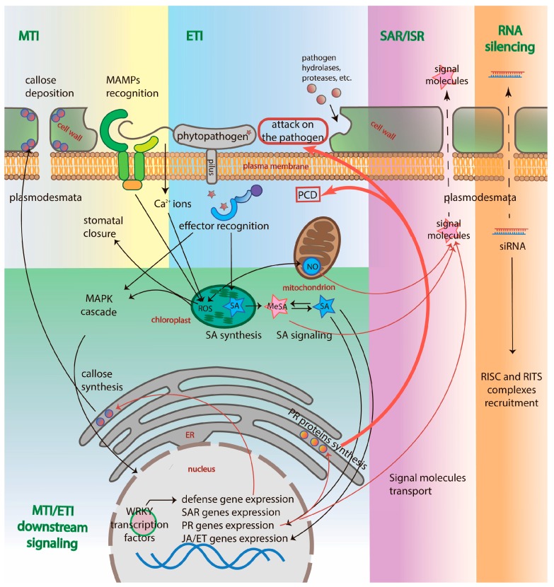
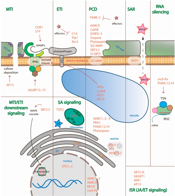
Plant defense is achieved mainly through the induction of microbe-associated molecular patterns (MAMP)-triggered immunity (MTI), effector-triggered immunity (ETI), systemic acquired resistance (SAR), induced systemic resistance (ISR), and RNA silencing. Plant immunity is a highly complex phenomenon with its own unique features that have emerged as a result of the arms race between plants and pathogens. However, the regulation of these processes is the same for all living organisms, including plants, and is controlled by proteases. Different families of plant proteases are involved in every type of immunity: some of the proteases that are covered in this review participate in MTI, affecting stomatal closure and callose deposition. A large number of proteases act in the apoplast, contributing to ETI by managing extracellular defense. A vast majority of the endogenous proteases discussed in this review are associated with the programmed cell death (PCD) of the infected cells and exhibit caspase-like activities. The synthesis of signal molecules, such as salicylic acid, jasmonic acid, and ethylene, and their signaling pathways, are regulated by endogenous proteases that affect the induction of pathogenesis-related genes and SAR or ISR establishment. A number of proteases are associated with herbivore defense. In this review, we summarize the data concerning identified plant endogenous proteases, their effect on plant-pathogen interactions, their subcellular localization, and their functional properties, if available, and we attribute a role in the different types and stages of innate immunity for each of the proteases covered.

Keywords: ETI; ISR; MTI; RNA silencing; SAR; plant immunity; plant proteases.


Conflict of interest statement

The authors declare no conflict of interest.

Figures

Figure 1
Figure 1
Plant defense mechanisms. MTI is triggered by MAMPs, leading to the elevation of cytosolic calcium ions, ROS and RNI generation, callose deposition at plasmodesmata and stomatal closure [39]. Effectors trigger ETI through binding to R proteins (NB-LRRs) that induce the signaling of SA and the subsequent induction of PR, JA/ET-dependent ISR-related genes and SAR-related genes. PR proteins, such as chitinases, β-1,3-glucanases, proteases, etc., either directly attack the pathogen or induce the PCD of the infected cell. SA is converted into MeSA that is transported into distal parts of the plant, as well as other signal molecules, establishing SAR or ISR. siRNA are also transported into distal parts of the plant through plasmodesmata. Names of immune processes are colored green. The names of cellular compartments are colored red. Black arrows indicate the directions of the activated plant immunity signaling pathways; red arrows indicate the results of genes expression after immunity activation; red bold arrows point to the cell fate in response to the pathogen; dotted arrows indicate the transport of signaling molecules through plasmodesmata.
Figure 2
Figure 2
The domain architecture of proteases covered in the review from different protease families. Red letters represent catalytic amino acid residues. SP—signal peptide, P—prodomain, C-term—C-terminal domain or granulin domain (for family C1A proteases), LP—linker peptide, NLS—nuclear localization signal, TM—transmembrane region, regions in grey—low complexity regions.
Figure 3
Figure 3
Involvement of plant proteases in different immunity pathways and their subcellular localization. Proteases covered in the review are colored orange. Immune processes are colored green. The names of cellular compartments are colored blue. Red arrows point to the object of the protease action (hydrolysis of the substrate or influence on the process); T-like arrows imply inhibition of proteases by effectors; black arrow indicates the direction of the MAPK cascade action; dotted arrow indicates the transport of signaling molecules through plasmodesmata.

References

    1. Anderson J.P., Gleason C.A., Foley R.C., Thrall P.H., Burdon J.B., Singh K.B. Plants versus pathogens: An evolutionary arms race. Funct. Plant Biol. 2010;37:499–512. doi: 10.1071/FP09304. - DOI - PMC - PubMed
    1. Santori F.R. The immune system as a self-centered network of lymphocytes. Immunol. Lett. 2015;166:109–116. doi: 10.1016/j.imlet.2015.06.002. - DOI - PMC - PubMed
    1. Farber D.L., Netea M.G., Radbruch A., Rajewsky K., Zinkernagel R.M. Immunological memory: Lessons from the past and a look to the future. Nat. Rev. Immunol. 2016;16:124–128. doi: 10.1038/nri.2016.13. - DOI - PubMed
    1. Ronald P.C., Beutler B. Plant and animal sensors of conserved microbial signatures. Science. 2010;330:1061–1064. doi: 10.1126/science.1189468. - DOI - PubMed
    1. Muthamilarasan M., Prasad M. Plant innate immunity: An updated insight into defense mechanism. J. Biosci. 2013;38:433–449. doi: 10.1007/s12038-013-9302-2. - DOI - PubMed

Substances

LinkOut - more resources