

THREE-YEAR OUTCOMES IN A RANDOMIZED SINGLE-BLIND CONTROLLED TRIAL OF INTRAVITREAL RANIBIZUMAB AND ORAL SUPPLEMENTATION WITH DOCOSAHEXAENOIC ACID AND ANTIOXIDANTS FOR DIABETIC MACULAR EDEMA
- PMID: 29474306
- PMCID: PMC6553973
- DOI: 10.1097/IAE.0000000000002114
THREE-YEAR OUTCOMES IN A RANDOMIZED SINGLE-BLIND CONTROLLED TRIAL OF INTRAVITREAL RANIBIZUMAB AND ORAL SUPPLEMENTATION WITH DOCOSAHEXAENOIC ACID AND ANTIOXIDANTS FOR DIABETIC MACULAR EDEMA
Abstract
Purpose: To report 3-year results of a randomized single-blind controlled trial of intravitreal ranibizumab combined with oral docosahexaenoic acid (DHA) supplementation versus ranibizumab alone in patients with diabetic macular edema.
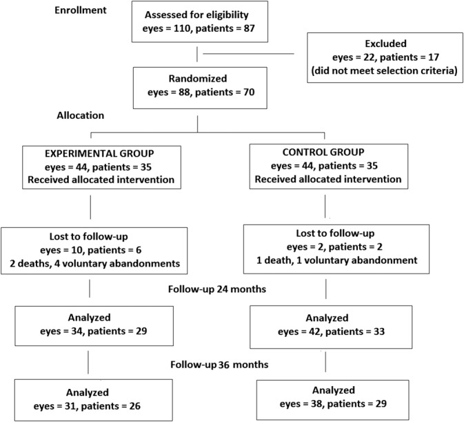
Methods: There were 26 patients (31 eyes) in the DHA group and 29 (38 eyes) in the control group. Ranibizumab (0.5 mg) was administered monthly for the first 4 months followed by a pro re nata (PRN) regimen. In the experimental group, patients received oral DHA supplementation (1,050 mg/day) (Brudyretina 1.5 g).
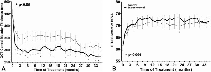
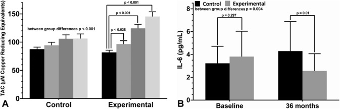
Results: At 36 months, mean decrease of central subfield macular thickness was higher in the DHA-supplementation group than in controls (275 ± 50 μm vs. 310 ± 97 μm) with significant differences at Months 25, 30, 33, and 34. Between-group differences in best-corrected visual acuity were not found, but the percentages of ETRDS gains >5 and >10 letters were higher in the DHA-supplementation group. Differences serum HbA1c, plasma total antioxidant capacity values, erythrocyte DHA content, and serum IL-6 levels were all significant in favor of the DHA-supplementation group.
Conclusion: The addition of a high-rich DHA dietary supplement to intravitreal ranibizumab was effective to achieve better sustained improvement of central subfield macular thickness outcomes after 3 years of follow-up as compared with intravitreal ranibizumab alone.
Conflict of interest statement
None of the authors has conflicting interests to disclose.
Figures
References
-
- Das A, McGuire PG, Rangasamy S. Diabetic macular edema: pathophysiology and novel therapeutic targets. Ophthalmology 2015;122:1375–1394. - PubMed
-
- Cai J, Boulton M. The pathogenesis of diabetic retinopathy: old concepts and new questions. Eye 2000;16:242–260. - PubMed
-
- SanGiovanni JP, Chew EY. The role of omega-3 long-chain polyunsaturated fatty acids in health and disease of the retina. Prog Retin Eye Res 2005;24:87–138. - PubMed
MeSH terms
Substances
LinkOut - more resources
Full Text Sources
Other Literature Sources
Medical
