. 2018 Feb 23;14(2):e1006913.
doi: 10.1371/journal.ppat.1006913. eCollection 2018 Feb.

Epitopes for neutralizing antibodies induced by HIV-1 envelope glycoprotein BG505 SOSIP trimers in rabbits and macaques

Affiliations

Epitopes for neutralizing antibodies induced by HIV-1 envelope glycoprotein BG505 SOSIP trimers in rabbits and macaques

P J Klasse et al. PLoS Pathog. .

Abstract

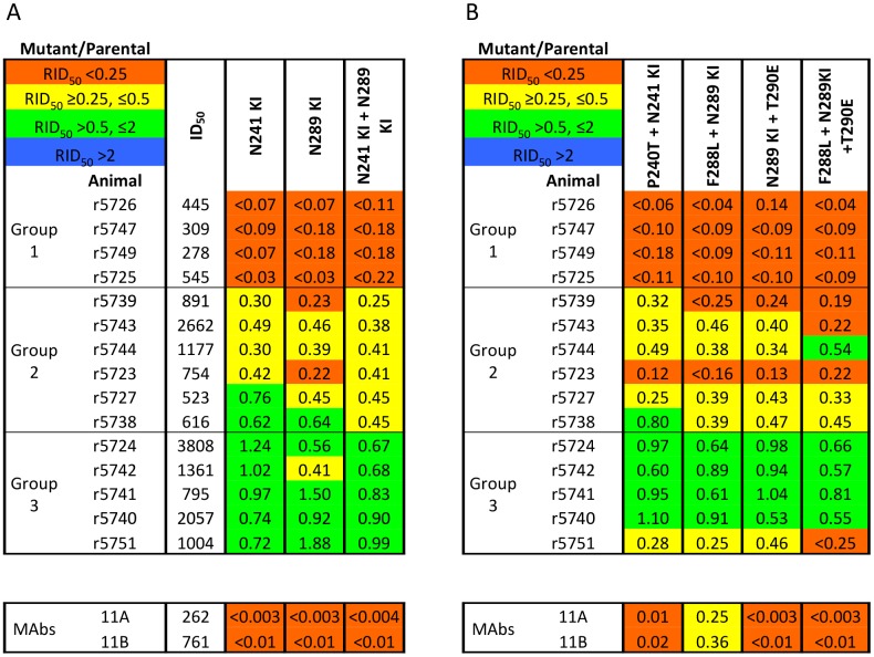
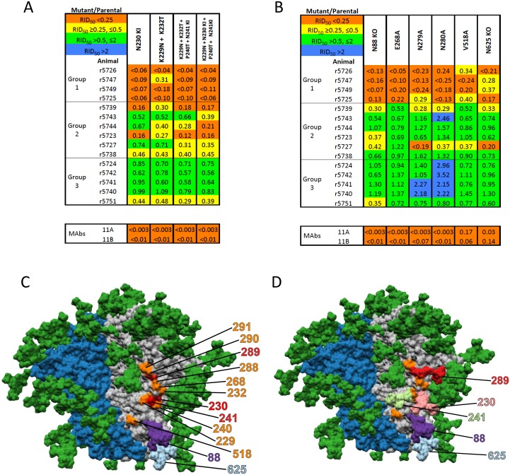
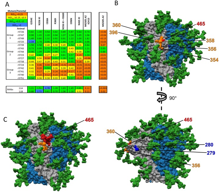
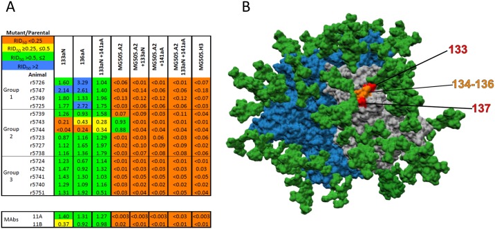
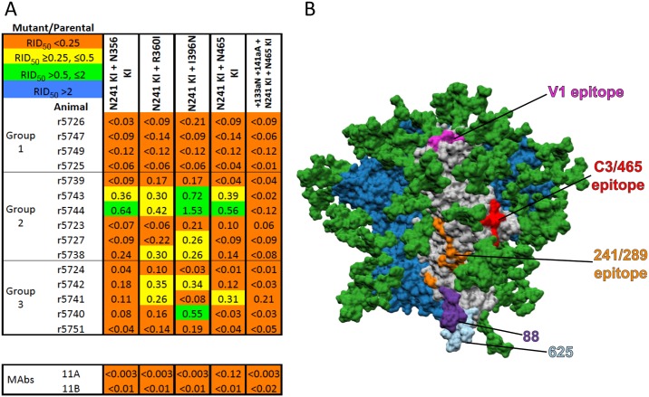
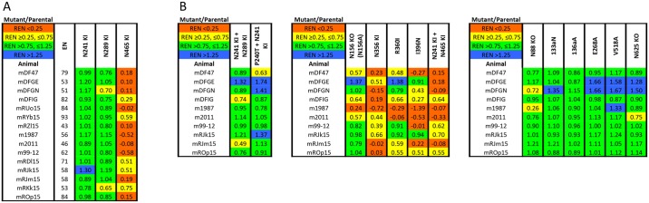
The native-like, soluble SOSIP.664 trimer based on the BG505 clade A env gene of HIV-1 is immunogenic in various animal species, of which the most studied are rabbits and rhesus macaques. The trimer induces autologous neutralizing antibodies (NAbs) consistently but at a wide range of titers and with incompletely determined specificities. A precise delineation of immunogenic neutralization epitopes on native-like trimers could help strategies to extend the NAb response to heterologous HIV-1 strains. One autologous NAb epitope on the BG505 Env trimer is known to involve residues lining a hole in the glycan shield that is blocked by adding a glycan at either residue 241 or 289. This glycan-hole epitope accounts for the NAb response of most trimer-immunized rabbits but not for that of a substantial subset. Here, we have used a large panel of mutant BG505 Env-pseudotyped viruses to define additional sites. A frequently immunogenic epitope in rabbits is blocked by adding a glycan at residue 465 near the junction of the gp120 V5 loop and β24 strand and is influenced by amino-acid changes in the structurally nearby C3 region. We name this new site the "C3/465 epitope". Of note is that the C3 region was under selection pressure in the infected infant from whom the BG505 virus was isolated. A third NAb epitope is located in the V1 region of gp120, although it is rarely immunogenic. In macaques, NAb responses induced by BG505 SOSIP trimers are more often directed at the C3/465 epitope than the 241/289-glycan hole.


Conflict of interest statement

The authors have declared that no competing interests exist.

Figures

Fig 1
Fig 1. A panel of rabbit sera with a spectrum of sensitivities to glycan knock-in mutations at positions 241 or 289 or both.
For each serum designated to the left, the ID50 values against each mutant pseudovirus relative to the BG505.T332N parental virus (RID50 = relative ID50, i.e., (ID50 against mutant)/(ID50 against parental)) are recorded, and then color-coded as outlined in the top-left key. Thus, mutations conferring insignificant, intermediate or strong neutralization resistance are in green, yellow or red, respectively. The Env mutations are summarized in the top row, with the complete sequence changes in the mutants listed in S1 Table. KI = glycan knock-in. Each tabulated value is the midpoint or median of ≥2 replicates. The sera are rank-ordered from the strongest to the weakest effect of the N241-KI/N289-KI mutations and divided into sub-groups: Group-1, complete neutralization resistance; Group-2, intermediate outcomes; Group-3, no effect on neutralization. A. The absolute inhibitory dilution factors against the parental virus (ID50) are given in the first data column. The single N241-KI and N289-KI mutants and the double N241-KI/N289-KI mutant had similar neutralization phenotypes within each of the three serum sub-groups. B. Non-glycan mutations that affect the lining of the 241/289-glycan hole, when combined with either the N241-KI or N289-KI changes, only sporadically increased resistance to the Group-2 or Group-3 sera.
Fig 2
Fig 2. Mutations with effects similar to those of the N241-KI and N289-KI changes on neutralization by rabbit sera.
The layout in panels A and B mirrors that of Fig 1A. A. The N230-KI mutation ablated neutralization by Group-1 sera and had an intermediate effect on Group-2 sera. Combining mutations around residue D230 with the N241-KI and the P240T mutations somewhat expanded the neutralization-reducing effects in Groups-1 and -2. B. Changes in C2 and the N88-KO or N625-KO mutations reduced neutralization by Group-1 sera, and hence resembled the N241-KI and N289-KI mutations. C. A surface-rendered model of the BG505 SOSIP.664 trimer seen from the side with the apex up. The peptidic surface on the protomer with marked residues is grey and that on the neighboring protomer is dark blue; the peptidic surface of the third protomer is not seen. The glycans are dark green. The D230 and S241 and N289 residues are highlighted in red and other residues involved in the epitope are in orange. The glycans at positions N88 and N625 are colored in purple and light blue, respectively. D. The model indicates the predicted orientations of the glycans present on the N230-KI (pink), N241-KI (light green) and N289-KI (red) mutants; the N88 and N625 glycans are also colored, as in panel-C. The model is based on PDB-5V8M.
Fig 3
Fig 3. Mutations identifying the C3/465 epitope as a new rabbit NAb epitope.
A. The layout is the same as in Fig 1. Specific mutations in C3 and V4, including N465-KI, conferred resistance to Group-3 and some Group-2 sera, but not to Group-1 sera; their effect was therefore the converse of the N241-KI and N289-KI changes. The K241S mutant of the MG505 cl.A2 virus had a very similar profile (compare the mutant with the parental clone shown in a separate column to the right, and see also Fig 4; all RID50 values are relative to the BG505.T332N parental virus). B. A surface-rendered model of the BG505 SOSIP.664 trimer seen from the side with the apex up. The peptidic surface on the protomer with marked residues is grey and those surfaces on the neighboring protomers are dark blue. The glycans are dark green, as in C. The residues G354, N356, I358, R360 and I396 are highlighted in orange and T465 in red; the upper and lower panels show the same model, but from two perspectives rotated by 90°. In the lower panel, residues N279 and N280 are also highlighted, in blue. C) Structural model indicating the predicted orientation of the glycan (in red) now present on the N465-KI mutant, with residues contributing to the C3/465 epitope in orange.
Fig 4
Fig 4. V1 mutations conferring neutralization resistance to two rabbit sera.
A. The layout is the same as in Fig 1. No serum neutralized the MG505 cl.H3 virus but two Group-2 sera did neutralize MG505 cl.A2. Inserting the two V1 residues (133aN and 136aA), present in cl.H3 but absent from cl.A2, into the parental BG505.T332N virus, alone or together, conferred resistance to only those two sera. B. An oblique view of the apex of the BG505 SOSIP.664 trimer with the peptidic surface of the protomer with marked residues colored in grey and those surfaces on the other two protomers colored in dark-blue. The glycans are dark green. The positions of V1 residues 133 and 137 are shown in red and 134–136 in orange.
Fig 5
Fig 5. Pan-resistance mutations that markedly affect nearly all rabbit sera.
A. The layout is the same as in Fig 1A. Combining the N241-KI mutation with the N465-KI or other point mutations that had correlated effects (e.g., those in C3) strongly reduced neutralization by all three sub-groups of sera. B. A surface-rendered model of the BG505 SOSIP.664 trimer seen from the side with the apex up. The peptidic surface on the protomer with marked residues is grey and those surfaces on the neighboring protomers are dark blue. Glycans are dark green. The 241/289-glycan hole epitope (orange), The C3/465 epitope (red) and the V1 epitope (magenta) are highlighted. The N88 and N625 glycans are also colored, in purple and light blue respectively.
Fig 6
Fig 6. Epitope mapping of autologous NAb responses in BG505 trimer-immunized macaques.
Macaque samples (analyzed as purified IgG) are arranged in the left-most column according to the studies they were obtained from. The effect on neutralization is relativized as the extent of inhibition of mutant over that of parental IgG at a concentration corresponding to a 1/50 dilution of serum (REN = relative extent of neutralization, color-coded as in the key at top-left). A. Fifteen macaque IgG samples were tested against mutants that most affected neutralization by rabbit sera. The extents of neutralization of the parental virus (EN, %) are given in the first data column. B. Ten macaque IgG preparations were tested against the N241-KI/N289-KI and the N241-KI/P240T double mutants as well as selected mutants involving C3, V1 and other residues.

Similar articles

Cited by

References

    1. Burton DR, Ahmed R, Barouch DH, Butera ST, Crotty S, Godzik A, et al. A Blueprint for HIV Vaccine Discovery. Cell Host Microbe. 2012;12(4):396–407. . - PMC - PubMed
    1. Escolano A, Dosenovic P, Nussenzweig MC. Progress toward active or passive HIV-1 vaccination. J Exp Med. 2017;214(1):3–16. doi: 10.1084/jem.20161765 . - DOI - PMC - PubMed
    1. Sanders RW, Moore JP. Native-like Env trimers as a platform for HIV-1 vaccine design. Immunol Rev. 2017;275(1):161–82. doi: 10.1111/imr.12481 . - DOI - PMC - PubMed
    1. Ward AB, Wilson IA. The HIV-1 envelope glycoprotein structure: nailing down a moving target. Immunol Rev. 2017;275(1):21–32. doi: 10.1111/imr.12507 . - DOI - PMC - PubMed
    1. Kwong PD. What Are the Most Powerful Immunogen Design Vaccine Strategies? A Structural Biologist’s Perspective. Cold Spring Harb Perspect Biol. 2017;9(11). doi: 10.1101/cshperspect.a029470 . - DOI - PMC - PubMed

Publication types

MeSH terms

LinkOut - more resources