. 2018 Feb 23;14(2):e1006888.
doi: 10.1371/journal.ppat.1006888. eCollection 2018 Feb.

Distinct susceptibility of HIV vaccine vector-induced CD4 T cells to HIV infection

Affiliations

Distinct susceptibility of HIV vaccine vector-induced CD4 T cells to HIV infection

Sarah Auclair et al. PLoS Pathog. .

Abstract

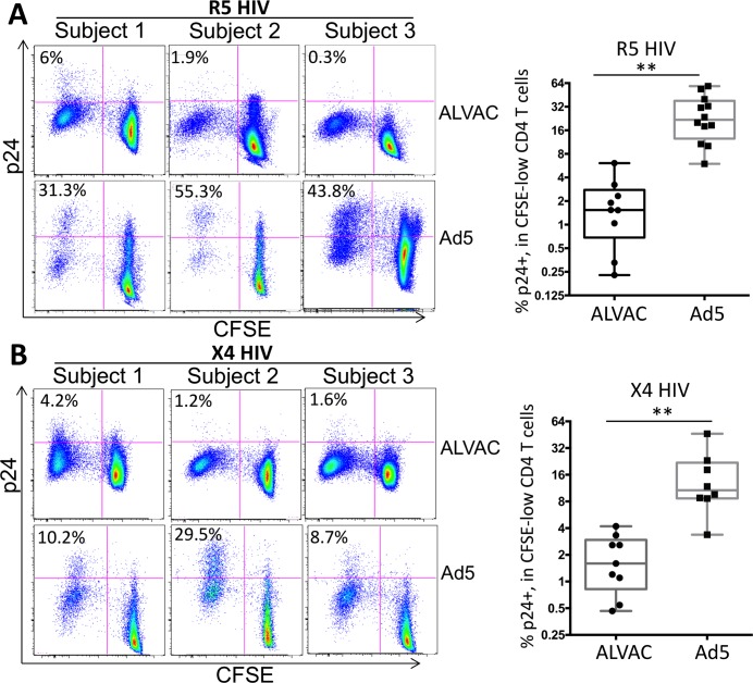
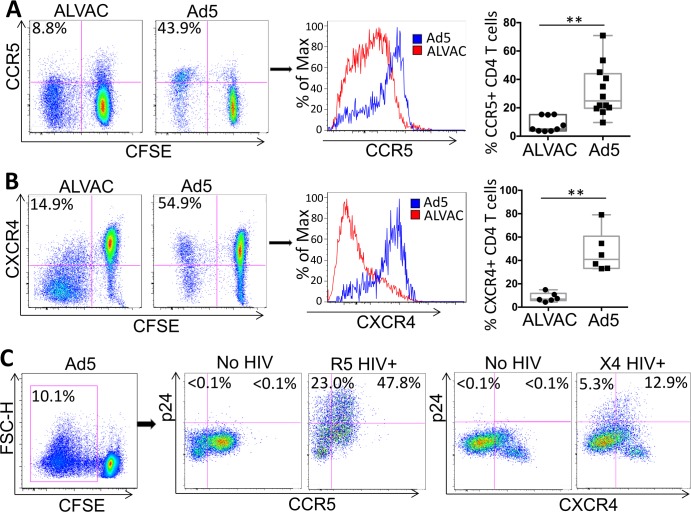
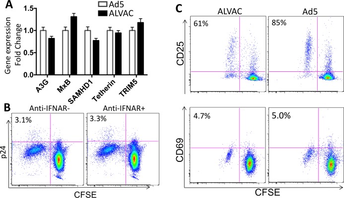
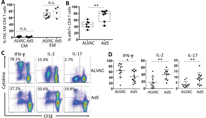
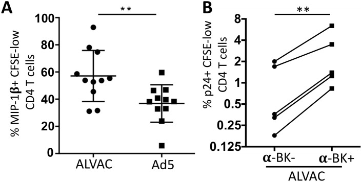
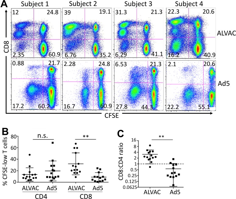
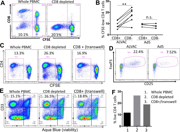
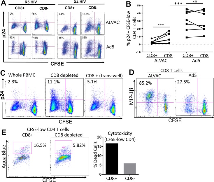
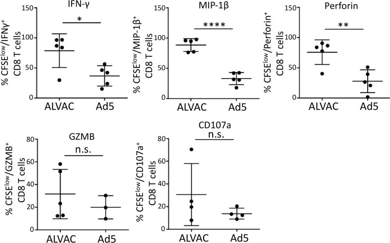
The concerns raised from adenovirus 5 (Ad5)-based HIV vaccine clinical trials, where excess HIV infections were observed in some vaccine recipients, have highlighted the importance of understanding host responses to vaccine vectors and the HIV susceptibility of vector-specific CD4 T cells in HIV vaccination. Our recent study reported that human Ad5-specific CD4 T cells induced by Ad5 vaccination (RV156A trial) are susceptible to HIV. Here we further investigated the HIV susceptibility of vector-specific CD4 T cells induced by ALVAC, a canarypox viral vector tested in the Thai trial RV144, as compared to Ad5 vector-specific CD4 T cells in the HVTN204 trial. We showed that while Ad5 vector-specific CD4 T cells were readily susceptible to HIV, ALVAC-specific CD4 T cells in RV144 PBMC were substantially less susceptible to both R5 and X4 HIV in vitro. The lower HIV susceptibility of ALVAC-specific CD4 T cells was associated with the reduced surface expression of HIV entry co-receptors CCR5 and CXCR4 on these cells. Phenotypic analyses identified that ALVAC-specific CD4 T cells displayed a strong Th1 phenotype, producing higher levels of IFN-γ and CCL4 (MIP-1β) but little IL-17. Of interest, ALVAC and Ad5 vectors induced distinct profiles of vector-specific CD8 vs. CD4 T-cell proliferative responses in PBMC, with ALVAC preferentially inducing CD8 T-cell proliferation, while Ad5 vector induced CD4 T-cell proliferation. Depletion of ALVAC-, but not Ad5-, induced CD8 T cells in PBMC led to a modest increase in HIV infection of vector-specific CD4 T cells, suggesting a role of ALVAC-specific CD8 T cells in protecting ALVAC-specific CD4 T cells from HIV. Taken together, our data provide strong evidence for distinct HIV susceptibility of CD4 T cells induced by different vaccine vectors and highlight the importance of better evaluating anti-vector responses in HIV vaccination.


Conflict of interest statement

The authors have declared that no competing interests exist.

Figures

Fig 1
Fig 1. ALVAC-specific CD4 T cells are markedly less susceptible to HIV infection in vitro than Ad5 vector-specific CD4 T cells.
PBMC collected from ALVAC- (RV144) or Ad5-vectored (HVTN204) HIV vaccine recipients were stained with CFSE and then re-stimulated with the recall vector antigen (ALVAC or Ad5) for three days before being infected with CCR5-tropic (US-1 strain) (A) or CXCR4-tropic (92/UG/029 strain) (B) HIV. HIV infection rate in vector-specific CD4 T cells was determined using flow cytometry to measure p24 expression 3 days post infection and expressed as the percentage of p24+ CFSE-low CD4 T cells. Representative flow cytometry plots shown at left are gated on CD3+CD8- CD4 T cells. Statistical analysis was performed using an unpaired Student’s t test. *p ≤ 0.05, **p ≤ 0.01.
Fig 2
Fig 2. ALVAC vector-specific CD4 T cells express lower levels of the HIV co-receptors CCR5 and CXCR4 than Ad5 vector-specific CD4 T cells.
PBMC of RV144 and HVTN204 vaccine recipients were stained with CFSE and stimulated with vector (ALVAC or Ad5) for 6 days. Surface expression of CCR5 (A) and CXCR4 (B) was measured by flow cytometry. Representative flow cytometry dot plots (left; gated on CD3+CD8- CD4 T cells) and histogram for co-receptor expression on ALVAC- and Ad5 vector-specific CD4 T cells are shown. Comparison of % CCR5+ or CXCR4+ vector-specific CD4 T cells from multiple subjects is shown (right). (C) HIV infection in co-receptor+ vs. co-receptor-, Ad5-specific CD4 T cells. CFSE-low, Ad5-specific CD4 T cells were gated for analysis (left). HIV infection rate (% p24+) in CCR5+ vs. CCR5- Ad5-specific CD4 T cells infected with R5 HIV (middle) or in CXCR4+ vs CXCR4- Ad5-specific CD4 T cells infected with X4 HIV (right) were shown. For both R5 and X4, no HIV infection was included as control to set p24 staining gate. Statistical analysis was performed using an unpaired Student’s t test; *p ≤ 0.05, **p ≤ 0.01.
Fig 3
Fig 3. ALVAC- and Ad5-specific CD4 T cells show similar levels of innate antiviral gene expression and immune activation.
(A) Relative expression of innate antiviral genes in ALVAC- and Ad5-specific CD4 T cells. RV144 and HVTN204 PBMC were CFSE-labeled and vector stimulated as described above. On day 6 ALVAC- and Ad5-specific CD4 T cells were sorted from PBMC based on CFSE-low and subjected to quantitative PCR for analysis of gene expression. The results were shown as fold change of ALVAC relative to Ad5. (B) HIV infection of ALVAC-specific CD4 T cells in RV144 PBMC in the presence or absence of anti-human IFNAR antibody blockade (gated on CD3+CD8- CD4 T cells). Number in each plot shows %p24+ in CFSE-low CD4 T cells. (C) Surface expression of T-cell activation markers CD25 (top) and CD69 (bottom) on ALVAC- vs. Ad5-specific CD4 T cells 6 days after stimulation with the corresponding vector (gated on CD3+CD8- CD4 T cells). Number in each plot shows % CD25+ or % CD69+ in CFSE-low CD4 T cells.
Fig 4
Fig 4. Phenotypic characterization of ALVAC- and Ad5 vector-specific CD4 T cells.
PBMC from RV144 or HVTN204 vaccine recipients were stained with CFSE and re-stimulated with vector for 6 days. Phenotypes and cytokine profile of CFSE-low, vector-specific CD4 T cells were measured by flow cytometry. (A) Comparison for percent of central memory (CM: CCR7+CD45RO+) and effector memory (EM: CCR7-CD45R)+) subsets in CFSE-low, ALVAC- and Ad5 vector-specific CD4 T cells; (B) Comparison for α4β7+% in CFSE-low, ALVAC- and Ad5 vector-specific CD4 T cells; (C) Representative flow cytometric plots for cytokine expression (IFN-γ, IL-2, and IL-17) in CFSE-low, ALVAC-specific (top) or Ad5 vector-specific (bottom) CD4 T cells; (D) Comparison for cytokine expression in CFSE-low, vector-specific CD4 T cells (% cytokine+ CFSE-low) between ALVAC and Ad5 vector from multiple vaccine recipients (n = 11). n.s.: not significant, *p ≤ 0.05, **p ≤ 0.01.
Fig 5
Fig 5. ALVAC-specific CD4 T cells produce higher levels of MIP-1β than Ad5-specific CD4 T cells, which contributes partly to their lower susceptibility to in vitro HIV infection.
(A) MIP-1β expression in CFSE-low, vector-specific CD4 T cells was determined by intracellular cytokine staining and flow cytometric analysis as described above; results are expressed as % MIP-1β+ CFSE-low CD4 T cells (n = 11). (B) Impact of MIP-1β neutralization on HIV infection of ALVAC-specific CD4 T cells. PBMC were stained with CFSE and re-stimulated in vitro with ALVAC vector in the absence of presence of β-chemokine neutralizing antibodies (CCL3/4/5). 3 days after vector stimulation, PBMC were infected with R5 HIV, followed by measurement of HIV infection in vector-specific CD4 T cells (CFSE-low, CD4 T cells) on day 6 after initial vector stimulation. HIV infection was expressed as the percentage of p24+ in CFSE-low CD4 T cells (n = 4). *p ≤ 0.05, **p ≤ 0.01.
Fig 6
Fig 6. ALVAC elicits distinct profile of vector-specific CD8 vs. CD4 T-cell proliferative response compared to Ad5 vector.
PBMC were stained with CFSE and re-stimulated with the corresponding vector for 6 days. (A) Representative flow cytometry plots for PBMC of multiple subjects showing vector-induced CD8 vs. CD4 T-cell proliferative responses in PBMC of RV144 (top) and HVTN204 (bottom) vaccine recipients. (B) Comparison for vector-specific CD8 and CD4 T-cell proliferative responses (% CFSE-low) in PBMC of RV144 and HVTN204 after corresponding vector stimulation. (C) Ratio of vector-specific CD8/CD4 T-cell proliferation in RV144 (ALVAC) and HVTN204 (Ad5) PBMC. Statistical analysis was performed using an unpaired Student’s t test; n = 14. *p ≤ 0.05, **p ≤ 0.01, ***p ≤ 0.001.
Fig 7
Fig 7. ALVAC-induced CD8 T cells inhibit the expansion of autologous vector-specific CD4 T cells.
(A) CD8+ cells were depleted from PBMC of vaccine recipients using magnetic beads. CD8-depleted or whole PBMC were CFSE stained and re-stimulated with the appropriate vector for 6 days. Efficient CD8 depletion was verified by flow cytometry. Number in the bottom-left quadrant shows % CFSE-low, proliferating CD4 T cells in total CD4 T cells. (B) Comparison for vector-specific CD4 T cell proliferation (% CFSE-low) in PBMC with or without CD8 depletion (n = 7 for ALVAC; n = 4 for Ad5). (C) CD4 T-cell proliferation in RV144 PBMC 6 days after stimulation with ALVAC. Comparison of whole PBMC, CD8-depleted PBMC, and PBMC from which CD8 T cells were depleted and then added back to culture in trans-well (gated on CD3+ T cells). (D) CD25 and FoxP3 expression in ALVAC- versus Ad5-specific CD8 T cells 6 days after stimulation with the corresponding vector (gated on CD3+CD8+ CFSE-low T cells). (E) Flow cytometry plot and (F) bar graph showing CD4 T cell viability (% viable cells) in RV144 PBMC 3 days after stimulation with ALVAC (before significant T-cell proliferation occurs), as determined by Aqua Blue dye exclusion. Comparison of cell viability in whole PBMC, CD8-depleted PBMC, and PBMC from which CD8 T cells were depleted then added back to culture in trans-well. Statistical analysis was performed using an unpaired Student’s t test; n = 2 (Ad5) or 7 (ALVAC). n.s.: non-significant; *p ≤ 0.05, **p ≤ 0.01.
Fig 8
Fig 8. CD8 depletion increases HIV susceptibility of ALVAC-specific CD4 T cells.
(A) Representative flow cytometry plots showing HIV infection in CFSE-low, vector-specific CD4 T cells in whole (CD8+) or CD8-depleted (CD8-) PBMC. Whole and CD8-depleted PBMC were CFSE stained and stimulated with vector antigen for 3 days before being infected with CCR5- or CXCR4-tropic HIV. HIV infection rate was determined using flow cytometry to measure intracellular p24 and expressed as the percentage of p24+ in CFSE-low CD4 T cells. CD3+CD8- T cells were gated for analysis. (B) Comparison for HIV infection rates in CFSE-low vector-specific CD4 T cells (% p24+) in whole or CD8-depleted PBMC from multiple vaccine recipients. (C) HIV infection (% p24+) in ALVAC-specific CD4 T cells in whole PBMC, CD8-depleted PBMC or PBMC from which CD8 T cells have been depleted and then added back to culture in trans-well (gated on CD3+CD8- CD4 T cells). % p24+ in CFSE-low cells was shown. (D) MIP-1β expression in ALVAC- versus Ad5-specific CD8 T cells 6 days after stimulation with the corresponding vector (gated on CD3+CD8+ T cells). (E) Flow cytometry plot (left) and bar graph (right) showing the viability of ALVAC-specific CD4 T cells (based on Aqua Blue staining) 6 days after vector stimulation with or without CD8 T-cell depletion (gated on CD3+CD8- CD4 T cells). Statistical analysis was performed using an unpaired Student’s t test; *p ≤ 0.05, **p ≤ 0.01, ***p ≤ 0.001.
Fig 9
Fig 9. ALVAC-induced CD8 T cells manifest stronger antiviral and cytotoxic phenotype than Ad5 vector-induced CD8 T cells.
PBMC of vaccine recipients were stained with CFSE and then stimulated with vector antigen for 6 days, followed by brief PMA/Ionomycin re-stimulation (6 hours) for cytokine/effector molecule re-synthesis. Intracellular staining and flow cytometry were used to measure the production of IFN-γ, MIP-1β, perforin, granzyme B (GZMB), and CD107a; results are expressed as % cytokine+ in CFSE-low CD8 T cells. Statistical analysis was performed using an unpaired Student’s t test; n = 3–5. n.s.: not significant; *p ≤ 0.05, **p ≤ 0.01, ***p ≤ 0.001, ****p ≤ 0.0001.

Similar articles

Cited by

References

    1. UNAIDS. Global AIDS Update 2016 Available from: http://www.unaids.org/en/resources/documents/2016/Global-AIDS-update-2016.
    1. Barouch DH. Novel adenovirus vector-based vaccines for HIV-1. Curr Opin HIV AIDS. 2010;5(5):386–90. doi: 10.1097/COH.0b013e32833cfe4c ; PubMed Central PMCID: PMC2967414. - DOI - PMC - PubMed
    1. Pantaleo G, Esteban M, Jacobs B, Tartaglia J. Poxvirus vector-based HIV vaccines. Curr Opin HIV AIDS. 2010;5(5):391–6. doi: 10.1097/COH.0b013e32833d1e87 . - DOI - PubMed
    1. Franchini G, Gurunathan S, Baglyos L, Plotkin S, Tartaglia J. Poxvirus-based vaccine candidates for HIV: two decades of experience with special emphasis on canarypox vectors. Expert Rev Vaccines. 2004;3(4 Suppl):S75–88. . - PubMed
    1. Buchbinder SP, Mehrotra DV, Duerr A, Fitzgerald DW, Mogg R, Li D, et al. Efficacy assessment of a cell-mediated immunity HIV-1 vaccine (the Step Study): a double-blind, randomised, placebo-controlled, test-of-concept trial. Lancet. 2008;372(9653):1881–93. Epub 2008/11/13. doi: 10.1016/S0140-6736(08)61591-3 ; PubMed Central PMCID: PMCPMC2721012. - DOI - PMC - PubMed

Publication types

MeSH terms