. 2018 Feb 23;13(2):e0193372.
doi: 10.1371/journal.pone.0193372. eCollection 2018.

Clinical and genetic characterisation of dystrophin-deficient muscular dystrophy in a family of Miniature Poodle dogs

Affiliations

Clinical and genetic characterisation of dystrophin-deficient muscular dystrophy in a family of Miniature Poodle dogs

Lluís Sánchez et al. PLoS One. .

Abstract

Four full-sibling intact male Miniature Poodles were evaluated at 4-19 months of age. One was clinically normal and three were affected. All affected dogs were reluctant to exercise and had generalised muscle atrophy, a stiff gait and a markedly elevated serum creatine kinase activity. Two affected dogs also showed poor development, learning difficulties and episodes of abnormal behaviour. In these two dogs, investigations into forebrain structural and metabolic diseases were unremarkable; electromyography demonstrated fibrillation potentials and complex repetitive discharges in the infraspinatus, supraspinatus and epaxial muscles. Histopathological, immunohistochemical and immunoblotting analyses of muscle biopsies were consistent with dystrophin-deficient muscular dystrophy. DNA samples were obtained from all four full-sibling male Poodles, a healthy female littermate and the dam, which was clinically normal. Whole genome sequencing of one affected dog revealed a >5 Mb deletion on the X chromosome, encompassing the entire DMD gene. The exact deletion breakpoints could not be experimentally ascertained, but we confirmed that this region was deleted in all affected males, but not in the unaffected dogs. Quantitative polymerase chain reaction confirmed all three affected males were hemizygous for the mutant X chromosome, while the wildtype chromosome was observed in the unaffected male littermate. The female littermate and the dam were both heterozygous for the mutant chromosome. Forty-four Miniature Poodles from the general population were screened for the mutation and were homozygous for the wildtype chromosome. The finding represents a naturally-occurring mutation causing dystrophin-deficient muscular dystrophy in the dog.


Conflict of interest statement

Competing Interests: One of the authors (AdS) is employed by a commercial company, Dick White Referrals. This does not alter our adherence to PLOS ONE policies on sharing data and materials. The authors declare that no other competing interests exist.

Figures

Fig 1
Fig 1. Pedigree showing the dystrophin-deficient muscular dystrophy affected MP family.
The litter is made up of three affected males, one unaffected male and one unaffected female. Both parents are also unaffected. DNA samples were available from dogs 1–6.
Fig 2
Fig 2. Representative histopathology of muscle cryosections from the infraspinatus muscle of dog 1.
Multifocal areas of myofibre degeneration are characterised by scattered and groups of necrotic fibres undergoing phagocytosis (a) and large clusters of basophilic regenerating fibres (b) consistent with a dystrophic phenotype (H&E stain).
Fig 3
Fig 3. Immunofluorescent analysis of muscle biopsies for localisation of dystrophy associated proteins.
Immunofluorescent staining of cryosections from dog 1 (dystrophic) and a control dog using antibodies to laminin α2, the rod domain and carboxy terminus (C-terminus) of dystrophin, dysferlin, α- and β-sarcoglycans (αSG and βSG respectively) and utrophin (DRP2). An antibody against spectrin was used as a control for membrane integrity. Staining was absent for the rod domain and carboxy terminus of dystrophin, decreased for β-sarcoglycans and similar for laminin α2, dysferlin, α-sarcoglycans and spectrin. Utrophin was visualized along the sarcolemma in the dystrophic dog but not on the sarcolemma of the control muscle. Regeneration was robust as shown by the antibody against developmental myosin heavy chain (dMHC).
Fig 4
Fig 4. Western blotting of skeletal muscle from dystrophic dogs (dogs 1 and 2).
Shown are stainings for the rod (DYS1) and carboxy terminus (DYS2) of dystrophin in the affected and control dogs. Bands are visible at approximately 395 kDA for the control muscle, but are not detectable in the dystrophic dogs. β-actin was used as a loading control and bands staining at a molecular weight of approximately 43 kDa confirm similar amounts of loaded protein in dystrophic and control dogs.
Fig 5
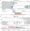
Fig 5. Breakpoint regions of the X chromosome deletion.
IGV display of the 5’ (A) and the 3’ breakpoint regions (B), in a DMD-affected and a control dog. The complete absence of reads in the DMD-affected dog is suggestive of a >5.6 Mb deletion (approximate coordinates X:26,238,000–31,869,800). Part of the reference sequence, CanFam3.1, is unknown (missing sequence in (A)). The specific regions targeted for amplification with primers are indicated, as are the highly homologous regions and the regions containing the deletion breakpoints (for the MP and GSHP, as described by VanBelzen et al.).
Fig 6
Fig 6. Quantitative PCR to genotype for the deletion.
A) IGV display of sequence reads for the DD-MD affected MPs and unaffected control at the qPCR loci. The DMD-del assay targets part of the dystrophin gene within the deletion, and the DMD-ref assay targets elsewhere on the X-chromosome. CanFam3.1 genes are represented by blue bars, and qPCR primers and probes are represented by red bars. B) Levels of DMD_del genomic DNA, normalised against DMD-ref, relative to a non-MP male dog (non-MP2). DMD_del is completely absent from the DD-MD cases, is present at levels of approximately 0.5x in dogs 5 and 6, and at level of approximately 1x in dog 4 and the non-MPs. Error bars represent standard deviation.

Similar articles

Cited by

References

    1. Koenig M, Monaco AP, Kunkel LM. The complete sequence of dystrophin predicts a rod-shaped cytoskeletal protein. Cell. 1988;53(2):219–28. . - PubMed
    1. Mendell JR, Shilling C, Leslie ND, Flanigan KM, al-Dahhak R, Gastier-Foster J, et al. Evidence-based path to newborn screening for Duchenne muscular dystrophy. Annals of neurology. 2012;71(3):304–13. doi: 10.1002/ana.23528 . - DOI - PubMed
    1. Caskey CT, Nussbaum RL, Cohan LC, Pollack L. Sporadic occurrence of Duchenne muscular dystrophy: evidence for new mutation. Clin Genet. 1980;18(5):329–41. . - PubMed
    1. Nowak KJ, Davies KE. Duchenne muscular dystrophy and dystrophin: pathogenesis and opportunities for treatment. EMBO Rep. 2004;5(9):872–6. doi: 10.1038/sj.embor.7400221 ; PubMed Central PMCID: PMCPMC1299132. - DOI - PMC - PubMed
    1. Hoffman EP, Brown RH Jr., Kunkel LM. Dystrophin: the protein product of the Duchenne muscular dystrophy locus. Cell. 1987;51(6):919–28. . - PubMed

Publication types

MeSH terms

LinkOut - more resources