Comparative Study
. 2018 Feb 23;13(2):e0193412.
doi: 10.1371/journal.pone.0193412. eCollection 2018.

The first in vivo multiparametric comparison of different radiation exposure biomarkers in human blood

Affiliations
Comparative Study

The first in vivo multiparametric comparison of different radiation exposure biomarkers in human blood

Ales Tichy et al. PLoS One. .

Abstract

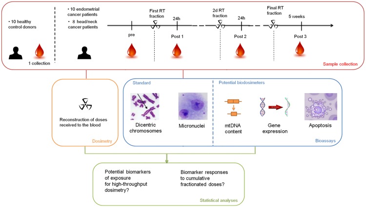
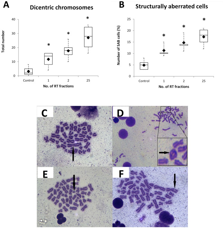
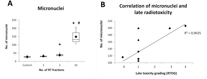
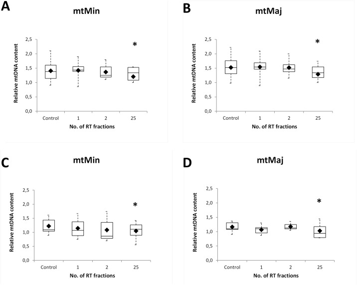
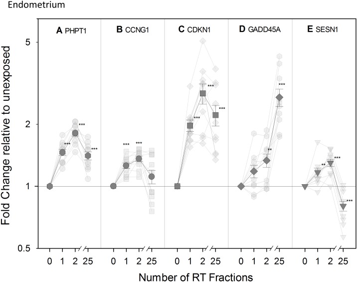
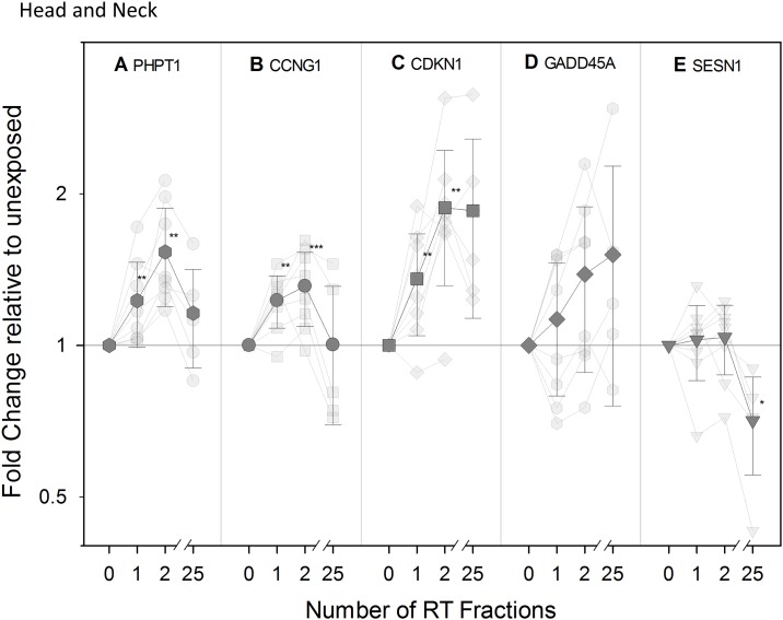
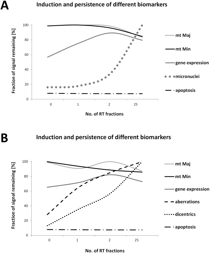
The increasing risk of acute large-scale radiological/nuclear exposures of population underlines the necessity of developing new, rapid and high throughput biodosimetric tools for estimation of received dose and initial triage. We aimed to compare the induction and persistence of different radiation exposure biomarkers in human peripheral blood in vivo. Blood samples of patients with indicated radiotherapy (RT) undergoing partial body irradiation (PBI) were obtained soon before the first treatment and then after 24 h, 48 h, and 5 weeks; i.e. after 1, 2, and 25 fractionated RT procedures. We collected circulating peripheral blood from ten patients with tumor of endometrium (1.8 Gy per fraction) and eight patients with tumor of head and neck (2.0-2.121 Gy per fraction). Incidence of dicentrics and micronuclei was monitored as well as determination of apoptosis and the transcription level of selected radiation-responsive genes. Since mitochondrial DNA (mtDNA) has been reported to be a potential indicator of radiation damage in vitro, we also assessed mtDNA content and deletions by novel multiplex quantitative PCR. Cytogenetic data confirmed linear dose-dependent increase in dicentrics (p < 0.01) and micronuclei (p < 0.001) in peripheral blood mononuclear cells after PBI. Significant up-regulations of five previously identified transcriptional biomarkers of radiation exposure (PHPT1, CCNG1, CDKN1A, GADD45, and SESN1) were also found (p < 0.01). No statistical change in mtDNA deletion levels was detected; however, our data indicate that the total mtDNA content decreased with increasing number of RT fractions. Interestingly, the number of micronuclei appears to correlate with late radiation toxicity (r2 = 0.9025) in endometrial patients suggesting the possibility of predicting the severity of RT-related toxicity by monitoring this parameter. Overall, these data represent, to our best knowledge, the first study providing a multiparametric comparison of radiation biomarkers in human blood in vivo, which have potential for improving biological dosimetry.


Conflict of interest statement

Competing Interests: The authors have declared that no competing interests exist.

Figures

Fig 1
Fig 1. Experimental design.
A scheme of blood sampling and subsequent analyses performed.
Fig 2
Fig 2. Chromosomal changes in PBMCs of head and neck patients.
Number of dicentric chromosomes (A) and structurally aberrant cells (B) was determined and both parameters increase with the number of RT fractions. *, statistically significant difference versus Control group (p < 0.01). Representative figures of dicentric chromosome (C) and structurally aberant cells—ring chromosome (D), double fragment (E) and break (F) are shown.
Fig 3
Fig 3. Incidence of micronuclei in PBMCs of endometrial patients (A) and their relation to late radiotoxicity (B).
Number of MN increased with the number of RT fractions. BN, binucleated cells; *, statistically significant difference of the 2nd and 25th RT fraction versus Control group (p < 0.001); #, statistically significant difference of the 25th RT fraction versus 2nd RT fraction (p < 0.001).
Fig 4
Fig 4. The relative proportion of early apoptotic, late apoptotic, and necrotic cells.
The relative proportions of early apoptotic (Annexin V-positive/propidium iodide-negative), late apoptotic (Annexin V-positive/propidium iodide-positive), and necrotic cells (Annexin V-negative/propidium iodide-positive) were determined (all combined set as 100%) in PBMC, granulocytes, and lymphocytes populations of endometrial (A) and head and neck patients (B). None of them showed a statistically significant change, but the necrotic population in head and neck patients after 25 RT fractions. See S1 Fig for gating strategy. *, statistically significant difference of the 25th RT fraction versus Control group (p < 0.05).
Fig 5
Fig 5. The amount of mtDNA content.
mtDNA was quantified in the minor arc (mtMin) and in the major arc (mtMaj) site. The data indicate the relative decrease in mtDNA content over the time in both groups of the endometrial (A) and (B) and head and neck (C) and (D) patients. Upper and lower quartiles with median are shown. Black rhombs represent arithmetic mean; *, statistically significant difference between 25th RT fraction and Control group (p < 0.05).
Fig 6
Fig 6. Transcriptional modification of expression of radiation-responsive genes in endometrial patients.
Transparent symbols represent QRT-PCR fold change in expression of the genes PHPT (A), CCNG1 (B), CDKN1A (C), GADD45 (D), and SESN1 (E) for ten individual endometrium cancer patients before (0) and after 1, 2, and 25 fractions of radiotherapy (RT). The mean (± SEM) is shown as non-transparent symbols. Gene expression is given as fold change relative to unexposed Control sample set at 1 (normalized against the housekeeping gene, HPRT). **, statistically significant difference versus Control group (p < 0.01); ***, statistically significant difference versus Control group (p < 0.005).
Fig 7
Fig 7. Transcriptional modification of expression of radiation-responsive genes in head and neck patients.
Transparent symbols represent QRT-PCR fold change in expression of the genes PHPT (A), CCNG1 (B), CDKN1A (C), GADD45 (D), and SESN1 (E) for eight head and neck cancer patients before (0) and after 1, 2, and 25 fractions of radiotherapy (RT). The mean (± SEM) is shown as non-transparent symbols. Gene expression is given as fold change relative to unexposed Control sample set at 1 (normalized against the housekeeping gene, HPRT). **, statistically significant difference versus Control group (p < 0.01); ***, statistically significant difference versus Control group (p < 0.005). For GADD45 p-trend was applied (r2 = 0.980).
Fig 8
Fig 8. Induction and persistence of different biomarkers.
Graphs showing the induction and temporal persistence of the biomarkers studied in endometrial (A) and head and neck patients (B). In order to compare all the parameters studied, the highest value detected was arbitrary set as 100%. Established cytogenetic markers and mtDNA content are suitable for late time points after irradiation while the new biomarkers as radiation-induced genes are more informative atearly time-points after radiation exposure. mt Maj, data from mtDNA content analysis of mtMajArch; mt Min, data from mtDNA content analysis of mtMinArch; gene expression, data from MQRT-PCR (average of five genes); micronuclei, data from micronuclei assay; aberrations, data from structural aberration analyses; dicentrics, data from dicentric chromosomes assay; apoptosis, data from flow-cytometric detection of apoptotic cells in lymphocyte population (percentage of apoptotic cells versus healthy-intact cells set as 100%).

References

    1. Blakely WF, Salter CA, Prasanna PGS. Early-response biological dosimetry—recommended countermeasure enhancements for mass-casualty radiological incidents and terrorism. Health Phys. 2005;89: 494–504. - PubMed
    1. Chaudhry MA. Biomarkers for human radiation exposure. J Biomed Sci. 2008;15: 557–563. doi: 10.1007/s11373-008-9253-z - DOI - PubMed
    1. Rothkamm K, Beinke C, Romm H, Badie C, Balagurunathan Y, Barnard S, et al. Comparison of established and emerging biodosimetry assays. Radiat Res. 2013;180: 111–119. doi: 10.1667/RR3231.1 - DOI - PMC - PubMed
    1. Kabacik S, Mackay A, Tamber N, Manning G, Finnon P, Paillier F, et al. Gene expression following ionising radiation: identification of biomarkers for dose estimation and prediction of individual response. Int J Radiat Biol. 2011;87: 115–129. doi: 10.3109/09553002.2010.519424 - DOI - PubMed
    1. Manning G, Kabacik S, Finnon P, Bouffler S, Badie C. High and low dose responses of transcriptional biomarkers in ex vivo X-irradiated human blood. Int J Radiat Biol. 2013;89: 512–522. doi: 10.3109/09553002.2013.769694 - DOI - PubMed

Publication types

MeSH terms

LinkOut - more resources