Review
. 2018 Mar;35(3):401-409.
doi: 10.1111/echo.13845. Epub 2018 Feb 23.

Contrast - in cardiac magnetic resonance imaging

Affiliations
Review

Contrast - in cardiac magnetic resonance imaging

Nirav Patel et al. Echocardiography. 2018 Mar.

Abstract

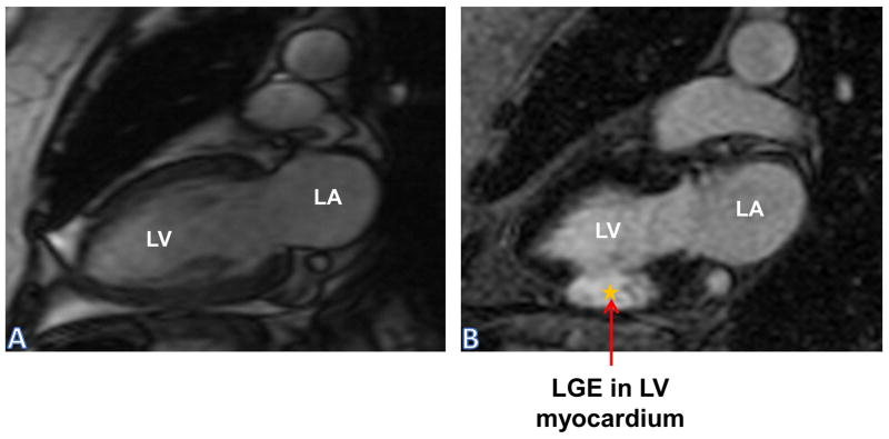
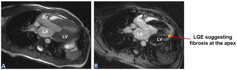
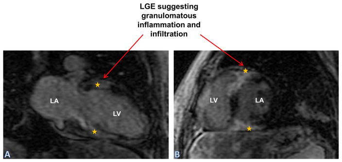
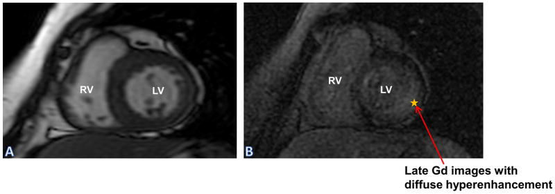
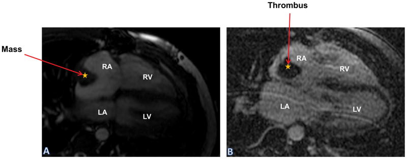
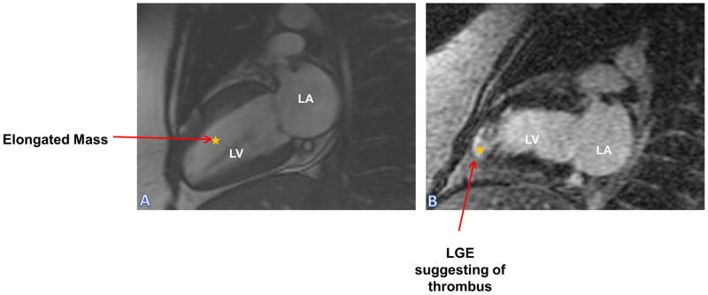
The utility and role of cardiac magnetic resonance (CMR) as a non-invasive diagnostic imaging modality has been well recognized in the field of cardiovascular disease. Use of late gadolinium enhancement (LGE) has further enhanced CMR's ability to determine structural, functional, and prognostic information in various cardiovascular diseases. The delivery and distribution of gadolinium as an extracellular agent allows the detection of edema, fibrosis, and infiltration in the myocardium. The pattern of LGE in cardiomyopathies enables us to distinguish among various disease processes non-invasively. Additionally, in patients with hypertrophic cardiomyopathy and sudden cardiac death, it helps in decision making in regards to use of implantable cardioverter defibrillator. In the evaluation of cardiac masses, LGE-CMR can often times differentiate between neoplastic and non-neoplastic conditions. In this review, we will discuss the various aspects of gadolinium-based contrast agents, and its application in CMR.

Keywords: cardiac magnetic resonance imaging; contrast imaging.


Figures

Figure 1
Figure 1
Coronary Artery Disease: Panel (A) SSFP sequence: Two chamber view showing left atrium and left ventricle shows normal thickness of inferior wall; Panel (B) LGE sequence showing areas of transmural hyper enhancement in mid-inferior wall suggesting infarction and non-viable myocardium. LA: Left Atrium; LGE: Late Gadolinium Enhancement; LV: Left Ventricle; SSFP: Steady State Free Precision
Figure 2
Figure 2
Hypertrophic Cardiomyopathy: Panel (A) SSFP sequence: left ventricle outflow tract image showing mid cavitary obliteration; Panel (B) LGE sequence showing hyper enhancement at the apex, suggestive of fibrosis in patient with hypertrophic cardiomyopathy. Ao: Aorta; LA: Left Atrium; LGE: Late Gadolinium Enhancement; LV: Left Ventricle; SSFP: Steady State Free Precision
Figure 3
Figure 3
Sarcoidosis: Panel (A) Two chamber (LA and LV) and Panel (B) Short axis view of LA and LV- both show areas of hyper enhancement secondary to granulomatous inflammation and infiltration in basal segments. LA: Left Atrium; LGE: Late Gadolinium Enhancement; LV: Left Ventricle
Figure 4
Figure 4
Amyloidosis: Panel (A) SSFP sequence of short axis LV and RV shows moderate concentric hypertrophy; Panel (B) LGE sequence shows diffuse hyper enhancement in lateral wall. LGE: Late Gadolinium Enhancement; LV: Left Ventricle; RV: Right Ventricle; SSFP: Steady State Free Precision
Figure 5
Figure 5
Late Gadolinium Images of LV slices at Base, mid-LV and Apex. Non-circumferential patchy LGE pattern is seen at base and mid-LV. No LGE is seen at Apex. LGE: Late Gadolinium Enhancement; LV: Left Ventricle; QALE: Query Amyloid Late Enhancement
Figure 6
Figure 6
Myocarditis: LGE sequence shows hyper enhancement of entire septum, inferior and anterior wall consistent with myocarditis. Note, enhancement of subepicardium and sparing of subendocardium on inferior and anterior walls. LGE: Late Gadolinium Enhancement; LV: Left Ventricle; RV: Right Ventricle
Figure 7
Figure 7
Arrhythmogenic right ventricular cardiomyopathy: Panels (A) and (B) SSFP sequence of four chamber and short axis showing RV dilatation; Panel (C) LGE sequence shows hyper enhancement in RV wall secondary to fibro fatty infiltration. LA: Left Atrium; LGE: Late Gadolinium Enhancement; LV: Left Ventricle; RA: Right Atrium; RV: Right Ventricle; SSFP: Steady State Free Precision
Figure 8
Figure 8
Effusive/Constrictive pericarditis: Panel (A) SSFP sequence of four chamber showing pericardial effusion with thickened pericardium; Panel (B) LGE sequence shows enhanced pericardium surrounding loculated effusion. LA: Left Atrium; LGE: Late Gadolinium Enhancement; LV: Left Ventricle; RA: Right Atrium; RV: Right Ventricle
Figure 9
Figure 9
Mass/Thrombus: Panel (A) SSFP sequence of four chamber showing filling defect in RA concerning for a myxoma; Panel (B) LGE sequence shows no enhancement of that mass suggesting against tumor and more likely a thrombus. LA: Left Atrium; LGE: Late Gadolinium Enhancement; LV: Left Ventricle; RA: Right Atrium; RV: Right Ventricle; SSFP: Steady State Free Precision
Figure 10
Figure 10
Thrombus: Panel (A) SSFP sequence of two chamber showing an elongated mass in the LV; Panel (B) LGE sequence shows enhancement of the apex, suggesting infarction and subsequent long serpiginous thrombus formation. LA: Left Atrium; LGE: Late Gadolinium Enhancement; LV: Left Ventricle; SSFP: Steady State Free Precision

Similar articles

Cited by

References

    1. Karamitsos TD, Francis JM, Myerson S, et al. The role of cardiovascular magnetic resonance imaging in heart failure. J Am Coll Cardiol. 2009;54:1407–1424. - PubMed
    1. Kwon DH, Smedira NG, Rodriguez ER, et al. Cardiac magnetic resonance detection of myocardial scarring in hypertrophic cardiomyopathy: correlation with histopathology and prevalence of ventricular tachycardia. J Am Coll Cardiol. 2009;54:242–249. - PubMed
    1. Soriano CJ, Ridocci F, Estornell J, et al. Noninvasive diagnosis of coronary artery disease in patients with heart failure and systolic dysfunction of uncertain etiology, using late gadolinium-enhanced cardiovascular magnetic resonance. J Am Coll Cardiol. 2005;45:743–748. - PubMed
    1. Reiter T, Ritter O, Prince MR, et al. Minimizing risk of nephrogenic systemic fibrosis in cardiovascular magnetic resonance. J Cardiovasc Magn Reson. 2012;14:31. - PMC - PubMed
    1. Friedrich MG, Sechtem U, Schulz-Menger J, et al. Cardiovascular magnetic resonance in myocarditis: A JACC White Paper. J Am Coll Cardiol. 2009;53:1475–1487. - PMC - PubMed

LinkOut - more resources