. 2018 Feb 23;37(1):37.
doi: 10.1186/s13046-018-0689-3.

SOX2OT variant 7 contributes to the synergistic interaction between EGCG and Doxorubicin to kill osteosarcoma via autophagy and stemness inhibition

Affiliations

SOX2OT variant 7 contributes to the synergistic interaction between EGCG and Doxorubicin to kill osteosarcoma via autophagy and stemness inhibition

Wanchun Wang et al. J Exp Clin Cancer Res. .

Abstract

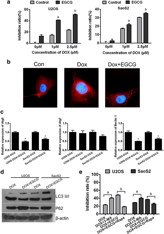
Background: Doxorubicin is the preferred chemotherapeuticdrug for osteosarcoma treatment of which clinical efficacy is limited because of its chemo-resistance and cardiac toxicity. It is necessary to develop the combination regimen with complementary molecular mechanisms to reduce the side effects and enhance sensitivity of Doxorubicin. EGCG is a polyphenol in green tea with antitumor bioactivity,which has been found that its combination with certain chemotherapeutic drugs could improve the antitumor efficiency.

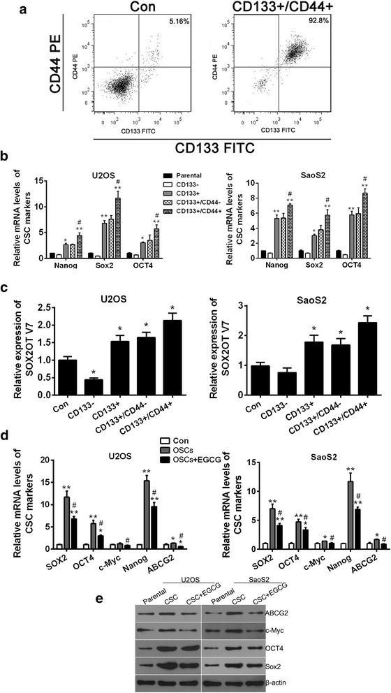
Methods: In this study, MTT assay was used to detect the cell growth inhibition The CD133+/CD44+ cells were isolated from U2OS and SaoS2 cell lines using magnetic-activated cell sorting and identified by flow cytometry analysis. qRT-PCR was used for determining the relative mRNA levels of key genes. Immunofluorescence was performed to evaluate the autophagy flux alterations. Self-renewal ability was accessed by sphere-forming assay. Tumorigenicity in nude mice was preformed to evaluate tumorigenicity in vivo.

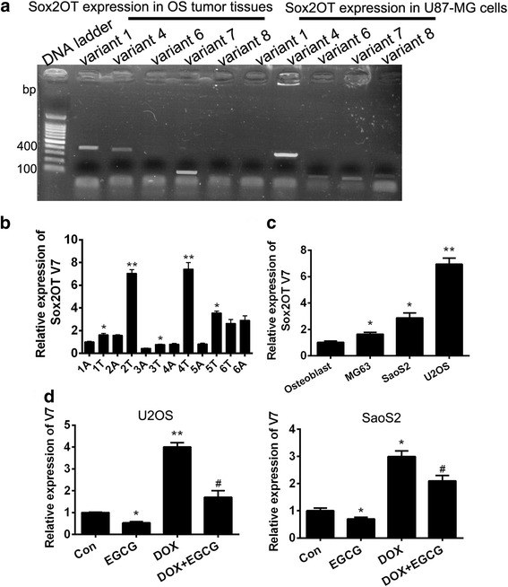
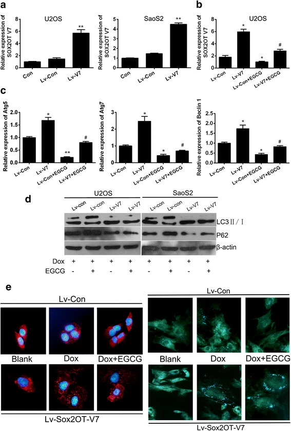
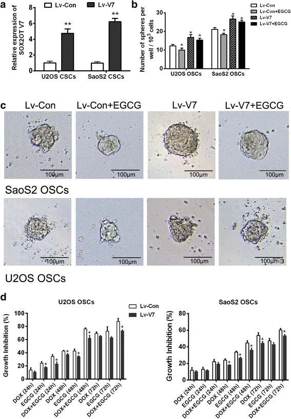
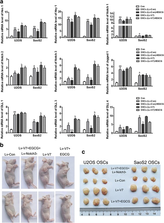
Results: We found that EGCG targeting LncRNA SOX2OT variant 7 produced synergistic effects with Doxorubicin on osteosarcoma cell growth inhibition. On the one hand, EGCG could reduce the Doxorubicin-induced pro-survival autophagy through decreasing SOX2OT variant 7 to improve the growth inhibition of Doxorubicin. On the other hand, EGCG could partially inactivate Notch3/DLL3 signaling cascade targeting SOX2OT variant 7 to reduce the stemness then abated drug-resistance of osteosarcoma cells.

Conclusions: This study will help to reveal the molecular mechanisms of synergistic effects of EGCG and Doxorubicin on OS chemotherapy and improve the clinical efficacy of chemotherapy as well as provide a basis for developing antitumor drugs targeting osteosarcoma stem cells.

Keywords: Autophagy; Cancer stem cells; Doxorubicin; EGCG; Notch3; Osteosarcoma; SOX2OT.


Conflict of interest statement

Ethics approval and consent to participate

Use of patient tissue samples for all experiments and the performance of animal experiments were approved by the Ethics Committee of the 2nd Xiangya Hospital of Central South University (2017 Ethical review No.S093).

Consent for publication

Not applicable

Competing interests

The authors declare that they have no competing interests.

Publisher’s Note

Springer Nature remains neutral with regard to jurisdictional claims in published maps and institutional affiliations.

Figures

Fig. 1
Fig. 1
Autophagy inhibition contributes to synergistic interaction between EGCG and Doxorubicin to kill the osteosarcoma cells. a Combination of EGCG and DOX synergistically facilitate antitumor effects in U2OS and SaoS2 cell lines. Columns, percentage of inhibition ratio; bars, SE. Data was from a representative of 5 independent studies. a p < 0.05 vs. control group treated without EGCG but only 1 μm Dox, b p < 0.05 vs. control group treated without EGCG but only 2.5 μm Dox. b Immunofluorescence analysis indicated that elevated LC3 fluorescent punctual signals were visualized in cells administrated with 2.5 mM DOX, while when EGCG was added, the LC3 fluorescence signal becomes weaker. c Exposed to 2.5 μM Dox with or without 20 μg/ml EGCG for 24 h, the expression levels of Atg5, Atg7 and Beclin1 in OS cells were determined at the mRNA levels by qRT-PCR. Results shown are representative of three independent experiments and error bars indicate SE. * p < 0.05 vs. DOX single treated group. d Cell lysates following DOX treatment with or without EGCG were subjected to western blotting. Protein ratios normalized to β-actin were used to quantify fold change. e Cells were under different treatment, then MTT assay was performed to detect the growth inhibition. Data was from a representative of 5 independent studies. a p < 0.05 vs. DOX treated group without 3-MA, b p < 0.05 vs. combined treated group without Rapa
Fig. 2
Fig. 2
Up-regulation of SOX2OT variant 7 in OS tumor tissues and cell lines. Specific expression of SOX2OTs (variants 1, 4, 6, 7, and 8) in OS tumor tissues. a The expression of SOX2OTs in OS tumor tissue mixture was checked by qRT-PCR. The U-87 cell line was used as positive control. b qRT-PCR was performed to detect the SOX2OT V7 in 6 pairs of OS tissues (No.1–6), A means adjacent and T means timorous. Results shown are representative of three independent experiments and error bars indicate SE. * p < 0.05 vs. relative adjacent tissue ** p < 0.01 vs. relative adjacent tissue. c qRT-PCR was performed to detect the mRNA level of SOX2OT V7 in 3 OS cell lines and primary osteoblast. Results shown are representative of three independent experiments and error bars indicate SE. * p < 0.05 vs. primary osteoblast group, ** p < 0.01 vs. primary osteoblast group. d qRT-PCR was performed to detect the mRNA level of SOX2OT V7 in U2OS and SaoS2 cells with different treatment. Results shown are representative of three independent experiments and error bars indicate SE. * p < 0.05 vs. control group, ** p < 0.01 vs. control group. # p < 0.05 vs. DOX single treated group
Fig. 3
Fig. 3
EGCG inhibited DOX-induced autophagy by targeting SOX2OT V7 partially. SOX2OT V7 gain-of-function cell models were constructed by lentivirus infection. a The relative mRNA levels of SOX2OT V7 in OS cells were checked by qRT-PCR. ** p < 0.01 vs. Lv-Con. b qRT-PCR was performed to detect the SOX2OT V7 in U2OS cells treated with or without EGCG. Results shown are representative of three independent experiments and error bars indicate SE. * p < 0.05 vs. Lv-con, # p < 0.05 vs. Lv-con treated with EGCG. c qRT-PCR was performed to detect the mRNA level of autophagy associated genes in V7 over-expressed U2OS cells. Results shown are representative of three independent experiments and error bars indicate SE. * p < 0.05 vs. Lv-con, # p < 0.05 vs. Lv-con treated with EGCG. d Western blots were performed to detect the expression of LC3 and P62 in V7 over-expressed U2OS and SaoS2 cells with different treatment. e LC3 immunofluorescence (the left half) and MDC staining (theright half), LC3 puncta distribution and MDC signals were visualized by confocal fluorescence microscopy, and representative images are shown. The images were captured at 400 x magnification
Fig. 4
Fig. 4
EGCG reduced the expression of CSC markers of osteosarcoma stem cell. a OSCs obtained from osteosarcoma cell line U2OS by CD133+/CD44+ immunomagnetic beads double positive screening and the isolated CD133+/CD44+ OSCs were identified by flow cytometry analysis. U2OS parental cells were set as Control (Con). b The relative mRNA levels of CSC markers Nanog, Sox2 and OCT4 in different bead-sorted cells were checked by qRT-PCR. * p < 0.05 vs. parental cells, ** p < 0.01 vs. parental cells, # p < 0.05 vs. CD133+ cells. c qRT-PCR was performed to detect the SOX2OT V7 in different bead-sorted cells. * p < 0.05 vs. parental cells (Con). d Relative mRNA levels of CSC markers Nanog, Sox2, OCT4, c-Myc and ABCG2 in OSCs treated with or without EGCG were detected by qRT-PCR. * p < 0.05 vs. parental cells (Con), ** p < 0.01 vs. parental cells (Con), # p < 0.05 vs. OSC. Results shown are representative of three independent experiments and error bars indicate SD. e Expression of CSC markers Nanog, Sox2, OCT4, c-Myc and ABCG2 in OSCs treated with or without EGCG were detected by western blots
Fig. 5
Fig. 5
SOX2OT V7 is one of the important targets of EGCG inhibiting OSCs. OSCs screened from U2OS and SaoS2 were infected with V7 overexpression lentivirus (Lv-V7) to generate gain-of-function OSC models of SOX2OT V7. a qRT-PCR was performed to detect the mRNA level of SOX2OT V7 in V7 overexpressed OSCs. ** p < 0.01 vs. control lentivirus infected OSCs (Lv-con). The sphere clone formation assay was performed to evaluate the self-renewal ability of OSCs. b 1 × 103 OSCs were inoculated in 96-well plate and number of spheres per well was calculated on day 14. * p < 0.05 vs. control lentivirus infected OSCs (Lv-con). c Representative pictures of a tumor spheres were taken on the day14, under 100 × magnification, the bar = 100 μm. d. MTT assay was performed to study the inhibitory effect of EGCG and DOX, alone or in combination on OSC growth. * p < 0.05 vs Lv-con OSCs which were treated under the same treatment condition
Fig. 6
Fig. 6
The OSC inhibitory effect of EGCG targeting SOX2OT V7 is partly achieved by restrain of Notch3 / DLL3 signaling. To illustrate whether the OSC inhibitory effect of EGCG targeting SOX2OT V7 is partly achieved by mediating of Notch signaling, relative mRNA levels of target genes, receptors and ligands of Notch signaling were detected using qRT-PCR in V7 overexpressed OSCs with or without EGCG treatment a.a p < 0.05 vs. parental cells (Con), aa p < 0.01 vs. parental cells (Con), bp < 0.05 vs. control lentivirus infected OSCs (Lv-con).b Typical pictures of nude mice with tumorigenicity which were under different treatments. c Typical pictures of stripped tumors derived from mice in different treatment groups

Similar articles

Cited by

References

    1. Ottaviani G, Robert RS, Huh WW, Palla S, Jaffe N. Sociooccupational and physical outcomes more than 20 years after the diagnosis of osteosarcoma in children and adolescents: limb salvage versus amputation. Cancer Biology Ther. 2013;119(20):3727–3736. - PMC - PubMed
    1. Huang J, Ni J, Liu K, Yu Y, Xie M, Kang R, Vernon P, Cao L. Tang D.HMGB1 promotes drug resistance in osteosarcoma. Cancer Res. 2012;72(1):230–238. doi: 10.1158/0008-5472.CAN-11-2001. - DOI - PubMed
    1. Chen L, Ye HL, Zhang G, Yao WM, Chen XZ, Zhang FC, Liang G. Autophagy inhibition contributes to the synergistic interaction between EGCG and doxorubicin to kill the hepatoma Hep3B cells. PLoS One. 2014;9(1):e85771. doi: 10.1371/journal.pone.0085771. - DOI - PMC - PubMed
    1. Zhang Z, Zhang Y, Lv J, Wang J. The survivin suppressant YM155 reverses doxorubicin resistance in osteosarcoma. Int J Clin Exp Med. 2015;8(10):18032–18040. - PMC - PubMed
    1. Kim HB, Lee SH, Um JH, Kim MJ, Hyun SK, Gong EJ, Oh WK, Kang CD, Kim SH. Sensitization of chemo-resistant human chronic myeloid leukemia stem-like cells to Hsp90 inhibitor by SIRT1 inhibition. Int J Biol Sci. 2015;11(8):923–934. doi: 10.7150/ijbs.10896. - DOI - PMC - PubMed

MeSH terms