. 2018 Feb:16:52-62.
doi: 10.1016/j.lssr.2017.11.002. Epub 2017 Nov 11.

Forces associated with launch into space do not impact bone fracture healing

Affiliations

Forces associated with launch into space do not impact bone fracture healing

Paul Childress et al. Life Sci Space Res (Amst). 2018 Feb.

Abstract

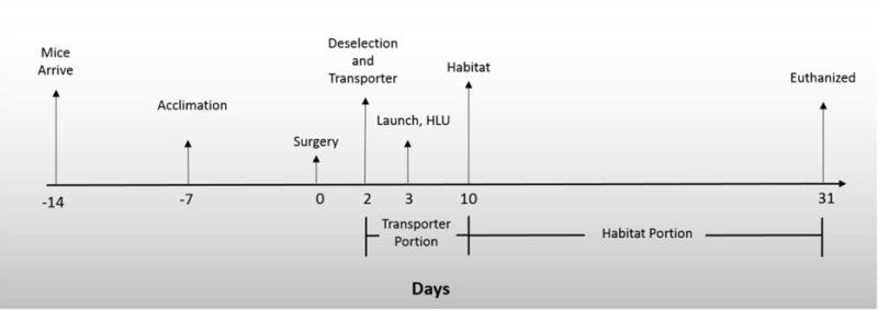
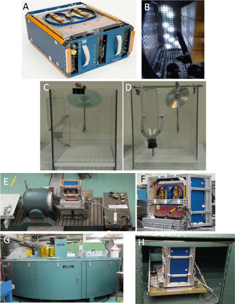
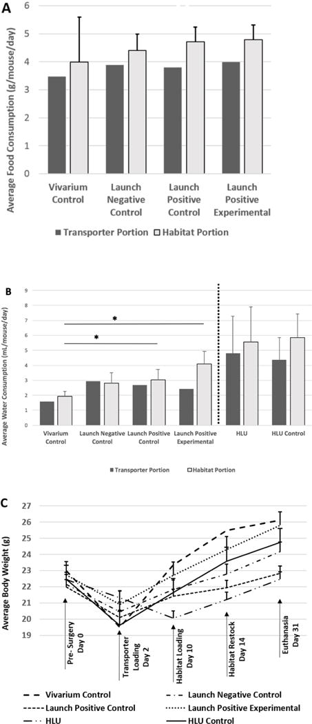
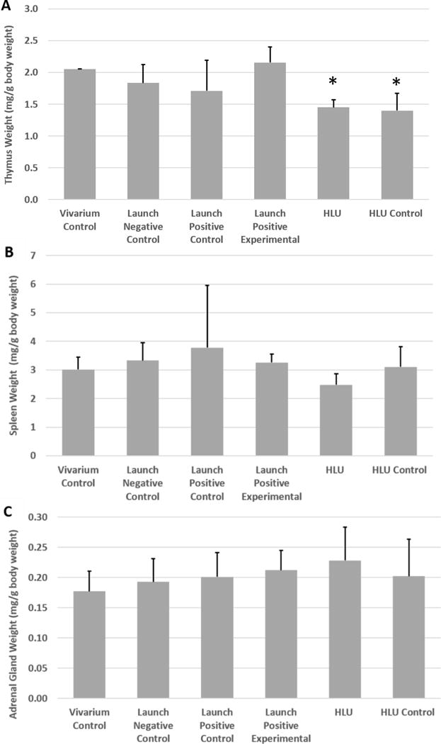
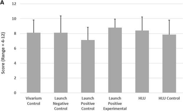
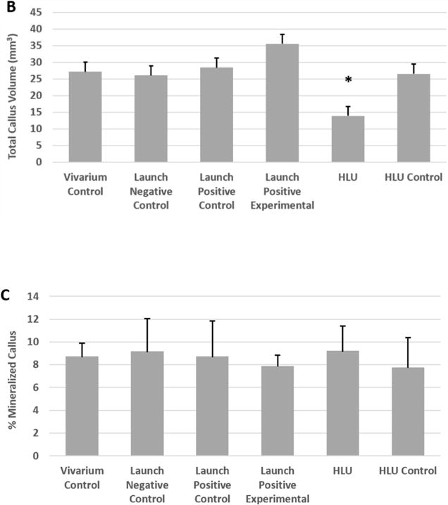
Segmental bone defects (SBDs) secondary to trauma invariably result in a prolonged recovery with an extended period of limited weight bearing on the affected limb. Soldiers sustaining blast injuries and civilians sustaining high energy trauma typify such a clinical scenario. These patients frequently sustain composite injuries with SBDs in concert with extensive soft tissue damage. For soft tissue injury resolution and skeletal reconstruction a patient may experience limited weight bearing for upwards of 6 months. Many small animal investigations have evaluated interventions for SBDs. While providing foundational information regarding the treatment of bone defects, these models do not simulate limited weight bearing conditions after injury. For example, mice ambulate immediately following anesthetic recovery, and in most cases are normally ambulating within 1-3 days post-surgery. Thus, investigations that combine disuse with bone healing may better test novel bone healing strategies. To remove weight bearing, we have designed a SBD rodent healing study in microgravity (µG) on the International Space Station (ISS) for the Rodent Research-4 (RR-4) Mission, which launched February 19, 2017 on SpaceX CRS-10 (Commercial Resupply Services). In preparation for this mission, we conducted an end-to-end mission simulation consisting of surgical infliction of SBD followed by launch simulation and hindlimb unloading (HLU) studies. In brief, a 2 mm defect was created in the femur of 10 week-old C57BL6/J male mice (n = 9-10/group). Three days after surgery, 6 groups of mice were treated as follows: 1) Vivarium Control (maintained continuously in standard cages); 2) Launch Negative Control (placed in the same spaceflight-like hardware as the Launch Positive Control group but were not subjected to launch simulation conditions); 3) Launch Positive Control (placed in spaceflight-like hardware and also subjected to vibration followed by centrifugation); 4) Launch Positive Experimental (identical to Launch Positive Control group, but placed in qualified spaceflight hardware); 5) Hindlimb Unloaded (HLU, were subjected to HLU immediately after launch simulation tests to simulate unloading in spaceflight); and 6) HLU Control (single housed in identical HLU cages but not suspended). Mice were euthanized 28 days after launch simulation and bone healing was examined via micro-Computed Tomography (µCT). These studies demonstrated that the mice post-surgery can tolerate launch conditions. Additionally, forces and vibrations associated with launch did not impact bone healing (p = .3). However, HLU resulted in a 52.5% reduction in total callus volume compared to HLU Controls (p = .0003). Taken together, these findings suggest that mice having a femoral SBD surgery tolerated the vibration and hypergravity associated with launch, and that launch simulation itself did not impact bone healing, but that the prolonged lack of weight bearing associated with HLU did impair bone healing. Based on these findings, we proceeded with testing the efficacy of FDA approved and novel SBD therapies using the unique spaceflight environment as a novel unloading model on SpaceX CRS-10.

Keywords: Fracture healing; Hindlimb unloading; Launch simulation; Spaceflight.


Figures

Figure 1
Figure 1. Experimental Timeline
Mice were purchased from Jackson Laboratories two weeks prior to surgery and placed in large N40 mouse cages. One week prior to surgery, raised wire floors were placed into N40 cages and mice began acclimation to spaceflight water Lixit hardware and spaceflight food bars. Two weeks after arrival mice underwent a segmental bone defect surgery. Two days after surgery some mice were placed in a Transporter. One day later some mice in Transporters were subjected to launch simulation and/or were placed in HLU cages and were either HLU or served as singly housed controls. One week later mice in Transporters were moved to Habitat caging. Thirty one days post-surgery all mice were euthanized and bones/tissue collected.
Figure 2
Figure 2. Hardware/Cages and Launch Simulation Testing Equipment
(A) Outside view of the Transporter, NASA developed cage for transport of mice from Earth to the ISS on the SpaceX Dragon. (B) Inside view of the Habitat, NASA developed cage used to house mice while on the ISS. (C) and (D) Hindlimb unloading (HLU) cages. Mice were suspended via tail by the clip with ambulation throughout the cage via a pulley system (Morey-Holton and Globus, 2002). (E) and (F) Vibration table for vibration testing (mimics SpaceX launch profile) and how the Transporter was secured to the vibration table. (G) and (H) 20G Centrifuge for g-force testing (mimics SpaceX launch profile) showing how the Transporter was secured on the centrifuge. Photos provided by NASA Ames Research Center.
Figure 3
Figure 3. Apparent Food and Water Depletion and Mouse Weight
(A) Apparent food and (B) water depletion were measured while the mice were housed in the different pieces of hardware. Mice were (C) weighed prior to surgery, 2 days after surgery (just before placement into Transporters), 1 week after launch simulation when mice were moved into Habitat cages, 2 weeks later when the Habitats were restocked with food and water, and at the time of euthanasia. Food and water consumption were expressed as values per mouse per day. Bars represent the mean ± SEM. There are no error bars associated with either food or water consumption during the Transporter phase because per standard protocol, the Transporter was only accessed after 7 days. Additionally, for the Habitat phase of the Launch Positive Experimental group, there were no water consumption error bars because the unit must be disassembled to take this measurement. No significant differences were detected between groups for food consumption during the Habitat phase. Significant differences were detected for water consumption between Vivarium and Launch Positive and Launch Positive Experimental groups (* indicates p<0.05) but not between Vivarium and Launch Negative groups (p>0.05). HLU and Vivarium/Habitat groups were not compared for water consumption due to different water delivery mechanisms (denoted by dotted line). However, HLU vs. HLU Controls were not significantly different from each other for water consumption. With regard to body weight, no significant differences were observed between the groups when using a repeated measures ANOVA followed by a Bonferroni post-hoc test (p<0.05).
Figure 4
Figure 4. Thymus, Spleen, and Adrenal Gland Weight
Weights of the (A) thymus, (B) spleen, and (C) adrenal gland were measured and normalized to body weight. Bars represent the mean ± SEM. No differences were detected between groups for spleen or adrenal gland weight; however, the two singly housed mouse groups (HLU and HLU Control) both experienced significantly lower thymus weights compared to all of the other groups (* indicates significant difference from all group housed cohorts, p<0.05).
Figure 5
Figure 5. Bone Healing. (A) Fracture Healing Score
Femurs were subjected to μCT imaging and then analyzed to assess bone healing. Fracture healing was assessed using the Radiographic Union Score for Tibial Fractures (RUST) method (but applied to femurs). This method assigns a numerical value ranging from 4 (not healed) to 12 (maximally healed) based on the assessment of union in the medial, lateral, anterior, and posterior cortices visible on anteroposterior and lateral μCT images. Bars represent the mean ± SEM. No significant differences were observed between the groups. (B) Callus Volume. Femurs were imaged by μCT and analyzed to assess the total callus volume in the separate test groups. Bars represent the mean ± SEM. *HLU resulted in a significant reduction in callus volume (p<0.05, compared to all other groups). (C) Mineralized Callus. Femurs were evaluated by μCT and the percentage of mineralized callus was measured. Bars represent the mean ± SEM. No significant differences were observed between the groups.

Similar articles

Cited by

References

    1. ABOU-ISMAIL UA, MAHBOUB HD. The effects of enriching laboratory cages using various physical structures on multiple measures of welfare in singly-housed rats. Lab Anim. 2011;45:145–53. - PubMed
    1. AGUIRRE JI, PLOTKIN LI, STEWART SA, WEINSTEIN RS, PARFITT AM, MANOLAGAS SC, BELLIDO T. Osteocyte apoptosis is induced by weightlessness in mice and precedes osteoclast recruitment and bone loss. J Bone Miner Res. 2006;21:605–15. - PubMed
    1. ANDROJNA C, MCCABE NP, CAVANAGH PR, MIDURA RJ. Effects of Spaceflight and Skeletal Unloading on Bone Fracture Healing. Clinical Reviews in Bone and Mineral Metabolism. 2012;10:61–70.
    1. BARTOS L, BRAIN PF. Physiological responses to social status and housing conditions in male mice subject to food competition tests. Bolletino di Zoologia. 1993;60:293–296.
    1. BLABER EA, DVOROCHKIN N, LEE C, ALWOOD JS, YOUSUF R, PIANETTA P, GLOBUS RK, BURNS BP, ALMEIDA EA. Microgravity induces pelvic bone loss through osteoclastic activity, osteocytic osteolysis, and osteoblastic cell cycle inhibition by CDKN1a/p21. PLoS One. 2013;8:e61372. - PMC - PubMed

LinkOut - more resources