

Interleukin-6/Stat3 signaling has an essential role in the host antimicrobial response to urinary tract infection
- PMID: 29475562
- PMCID: PMC5967986
- DOI: 10.1016/j.kint.2017.12.006
Interleukin-6/Stat3 signaling has an essential role in the host antimicrobial response to urinary tract infection
Abstract
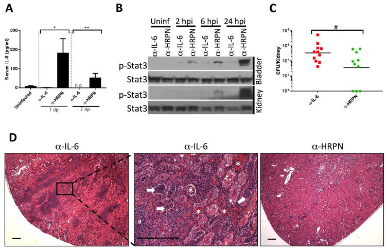
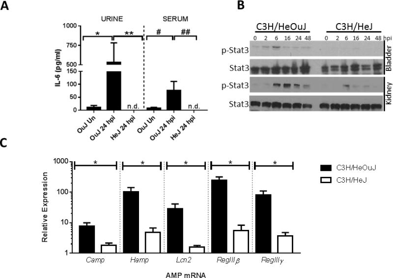
The signaling networks regulating antimicrobial activity during urinary tract infection (UTI) are incompletely understood. Interleukin-6 (IL-6) levels increase with UTI severity, but the specific contributions of IL-6 to host immunity against bacterial uropathogens are unknown. To clarify this we tested whether IL-6 activates the Stat3 transcription factor, to drive a program of antimicrobial peptide gene expression in infected urothelium during UTI. Transurethral inoculation of uropathogenic Escherichia coli led to IL-6 secretion, urothelial Stat3 phosphorylation, and activation of antimicrobial peptide transcription, in a Toll-like receptor 4-dependent manner in a murine model of cystitis. Recombinant IL-6 elicited Stat3 phosphorylation in primary urothelial cells in vitro, and systemic IL-6 administration promoted urothelial Stat3 phosphorylation and antimicrobial peptide expression in vivo. IL-6 deficiency led to decreased urothelial Stat3 phosphorylation and antimicrobial peptide mRNA expression following UTI, a finding mirrored by conditional Stat3 deletion. Deficiency in IL-6 or Stat3 was associated with increased formation of intracellular bacterial communities, and exogenous IL-6 reversed this phenotype in IL-6 knockout mice. Moreover, chronic IL-6 depletion led to increased renal bacterial burden and severe pyelonephritis in C3H/HeOuJ mice. Thus, IL-6/Stat3 signaling drives a transcriptional program of antimicrobial gene expression in infected urothelium, with key roles in limiting epithelial invasion and ascending infection.
Keywords: IL-6; Stat3; antimicrobial peptide; intracellular bacterial community; urinary tract infection; urothelium.
Copyright © 2018 International Society of Nephrology. Published by Elsevier Inc. All rights reserved.
Figures


Similar articles
-
A non-canonical autophagy-dependent role of the ATG16L1T300A variant in urothelial vesicular trafficking and uropathogenic Escherichia coli persistence.Autophagy. 2019 Mar;15(3):527-542. doi: 10.1080/15548627.2018.1535290. Epub 2018 Nov 8. Autophagy. 2019. PMID: 30335568 Free PMC article.
-
Expression and Significance of the HIP/PAP and RegIIIγ Antimicrobial Peptides during Mammalian Urinary Tract Infection.PLoS One. 2015 Dec 10;10(12):e0144024. doi: 10.1371/journal.pone.0144024. eCollection 2015. PLoS One. 2015. PMID: 26658437 Free PMC article.
-
Hepcidin as a Major Component of Renal Antibacterial Defenses against Uropathogenic Escherichia coli.J Am Soc Nephrol. 2016 Mar;27(3):835-46. doi: 10.1681/ASN.2014101035. Epub 2015 Aug 20. J Am Soc Nephrol. 2016. PMID: 26293821 Free PMC article.
-
Host-pathogen checkpoints and population bottlenecks in persistent and intracellular uropathogenic Escherichia coli bladder infection.FEMS Microbiol Rev. 2012 May;36(3):616-48. doi: 10.1111/j.1574-6976.2012.00339.x. FEMS Microbiol Rev. 2012. PMID: 22404313 Free PMC article. Review.
-
The bacteria and the host: a story of purinergic signaling in urinary tract infections.Am J Physiol Cell Physiol. 2021 Jul 1;321(1):C134-C146. doi: 10.1152/ajpcell.00054.2021. Epub 2021 May 12. Am J Physiol Cell Physiol. 2021. PMID: 33979212 Review.
Cited by
-
Molecular determinants of disease severity in urinary tract infection.Nat Rev Urol. 2021 Aug;18(8):468-486. doi: 10.1038/s41585-021-00477-x. Epub 2021 Jun 15. Nat Rev Urol. 2021. PMID: 34131331 Free PMC article. Review.
-
Rapid Bladder Interleukin-10 Synthesis in Response to Uropathogenic Escherichia coli Is Part of a Defense Strategy Triggered by the Major Bacterial Flagellar Filament FliC and Contingent on TLR5.mSphere. 2019 Nov 27;4(6):e00545-19. doi: 10.1128/mSphere.00545-19. mSphere. 2019. PMID: 31776239 Free PMC article.
-
PTEN modulates urinary tract infection susceptibility and shapes urothelial antibacterial defenses.Life Sci Alliance. 2025 Jul 23;8(10):e202503292. doi: 10.26508/lsa.202503292. Print 2025 Oct. Life Sci Alliance. 2025. PMID: 40701780 Free PMC article.
-
Immune defenses in the urinary tract.Trends Immunol. 2023 Sep;44(9):701-711. doi: 10.1016/j.it.2023.07.001. Epub 2023 Aug 15. Trends Immunol. 2023. PMID: 37591712 Free PMC article. Review.
-
The Potential Role of Persister Cells in Urinary Tract Infections.Curr Urol Rep. 2023 Nov;24(11):541-551. doi: 10.1007/s11934-023-01182-5. Epub 2023 Nov 1. Curr Urol Rep. 2023. PMID: 37907771 Review.
References
-
- Freedman AL. Urologic diseases in North America Project: trends in resource utilization for urinary tract infections in children. J Urol. 2005;173:949–954. - PubMed
Publication types
MeSH terms
Substances
Grants and funding
LinkOut - more resources
Full Text Sources
Other Literature Sources
Medical
Miscellaneous
