Review
. 2018 Mar-Apr;32(2):221-229.
doi: 10.21873/invivo.11228.

Non Lipomatous Benign Lesions Mimicking Soft-tissue Sarcomas: A Pictorial Essay

Affiliations
Review

Non Lipomatous Benign Lesions Mimicking Soft-tissue Sarcomas: A Pictorial Essay

Alessandro Coran et al. In Vivo. 2018 Mar-Apr.

Abstract

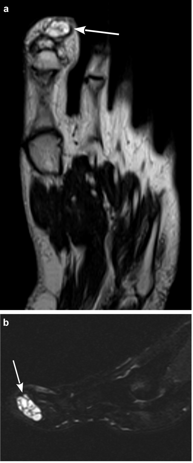
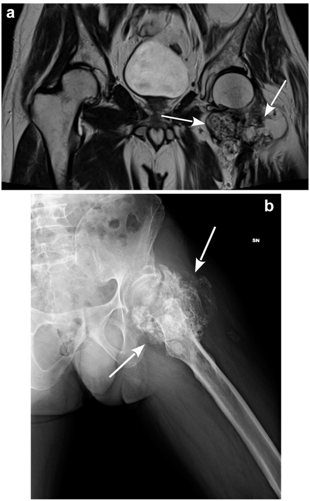
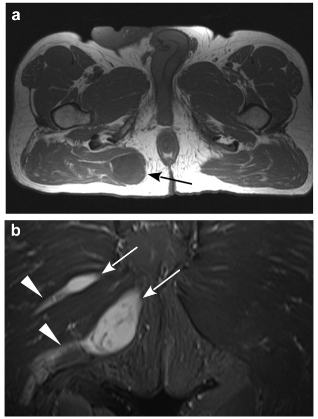
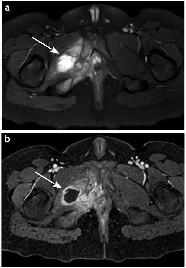
The incidental finding of soft tissue masses is a challenge for the radiologist. Benign and malignant lesions can be differentiated relying on patient history, symptoms and mostly with the help of imaging. Ultrasound (US), computed tomography (CT) and magnetic resonance imaging (MRI) become fundamental in order to distinguish these lesions but the radiologist needs to know the main characteristics of benign soft tissue masses and sarcomas. Herein, we present a pictorial review of lesions mimicking soft tissue sarcomas features.

Keywords: MRI; Sarcoma; mimics; review.


Figures

Figure 1
Figure 1. Synovial tendon cyst: a) Axial T2 weighted MRI scan showing a hyper-intense multi-lobulated mass of the first toe (white arrow); b)Sagittal STIR MRI scan highlighting the fluid content of the same lesion(white arrow). Synovial tendon cyst was found at histopathology.
Figure 2
Figure 2. Myositis ossificans: a) Coronal CT scan showing an inhomogeneous calcific mass (white arrow) of the left gluteus minimus; b) Coronal T2 weighted MRI scan shows an enlargement of the gluteus minimus with an inhomogeneous hyper-intense signal (white arrow) and focal areas of signal void. Myositis ossificans was proven with biopsy.
Figure 3
Figure 3. Synovial chondromatosis: a) Coronal T2 weighted MRI scan showing an inhomogeneous hyper-intense lesion (white arrows) with hypo-intense spots inside compatible with calcifications; b) X-ray showing diffusely calcific mass (white arrows). Synovial chondromatosis was found at histopathology.
Figure 4
Figure 4. Hemangioma: a) Axial T2 weighted MRI scan showing a hyperintense multi-lobulated mass of the tibialis anterior (with arrow) with multiple fluid-fluid levels; b) The same lesion (white arrow) on axial fatsaturated post-gadolinium T1 weighted MRI scan showing inhomogeneous contrast enhancement in the venous-phase and fluid-fluid levels. Later contrastographic phases demonstrated a progressive filling of the lesion (not shown). Hemangioma was found at histopathology.
Figure 5
Figure 5. Elastofibroma: a) Axial T2 weighted MRI scan showing hypointense subscapular formation (black arrow) between left serratus anterior muscle and chest wall, with hyper-intense lines consistent with fat tissue; b) Coronal STIR MRI image confirming the fat component of the lesion (white arrow). Elastofibroma was found at histopathology.
Figure 6
Figure 6. Nodular fasciitis: a) Coronal and b) sagittal T2 weighted MRI scan showing inhomogeneous hypo-intense nodule (white arrows) on the medial side of the plantar fascia, extended to subcutaneous tissue.Nodular fasciitis was found at histopathology.
Figure 7
Figure 7. Intramuscular myxoma: a) Axial T1 weighted MRI scan showing a hypo-intense nodule (black arrow) in the gluteus maximus muscle surrounded by a hyper-intense ring (fatty cap); b) Coronal STIR MRI scan showing the previously described lesion and another smaller nodule cranially (white arrows) both with homogeneous hyper-intense signal and peri-lesional hyper-intense offshoots (white arrowheads), consistent with leakage of myxomatous tissue. Intramuscular myxomas were found at histopathology.
Figure 8
Figure 8. Intramuscular schwannoma: a) Sagittal T2 weighted MRI scan showing an inhomogeneous lesion (white arrow) in the paravertebral muscles with a low intensity fibrous core (target sign); b) Sagittal T1 weighted MRI scan of the same area showing the hyper-intense fatty rim of the lesion (split fat sign). Intramuscular schwannoma was found at histopathology.
Figure 9
Figure 9. Muscular abscess: a) Axial STIR MRI scan showing a hyperintense lesion of the right external obturator muscle (white arrow) with surrounding edema; b) Axial T1 weighted MRI scan with fat saturation and after gadolinium injection showing the same area with a rim of peripheral contrast enhancement in venous phase (white arrow). Muscular abscess was diagnosed with needle biopsy.
Figure 10
Figure 10. Hematoma: a) Sagittal Proton Density weighted MRI scan showing an inhomogeneous hyper-intense mass (white arrow) of the posterior compartment of the right leg; b) The same lesion (white arrow) in a T1 weighted axial MRI scan with fat saturation presenting peripheral contrast enhancement. Imaging follow-up demonstrated the typical MRI progression of hematoma.
Figure 11
Figure 11. Arteriovenous malformation: a) Axial T2 weighted MRI scan showing a multi-lobulated lesion, between extensor carpi radialis muscles and brachioradialis muscle, that appears hyper-intense with flow-void inside high-flow vessels (white arrow); b) Axial T1 weighted MRI scan with fat signal suppression showing the same lesion with strong contrast enhancement of the abovementioned vessels (white arrow). Diagnosis of arteriovenous malformation was confirmed with both imaging and histopathology features.
Figure 12
Figure 12. Morton’s Neuroma: Axial T2 weighted MRI scan showing a fusiform lesion that appears hypo-intense compared to fat and hyperintense compared to muscles (white arrow), located in the third intermetatarsal space. Diagnosis of Morton’s Neuroma was confirmed combining imaging and clinical features.

Similar articles

Cited by

References

    1. Bridge JA, Hogendoorn P, Bridge JA, Fletcher CD. WHO classification of tumours of soft tissue and bone. International Agency for Research on Cancer. 2013
    1. McKenzie G, Raby N, Ritchie D. Non-neoplastic soft-tissue masses. Br J Radiol. 2009;82(981):775–785. - PubMed
    1. Balik MS, Erkut A, Guvercin Y, Bedir R. Synovial Sarcoma Mimicking Myositis Ossificans. Knee Surg Relat Res. 2016;28(3):249–252. - PMC - PubMed
    1. Murphey MD, Vidal JA, Fanburg-Smith JC, Gajewski DA. Imaging of synovial chondromatosis with radiologic-pathologic correlation. Radiographics. 2007;27(5):1465–1488. - PubMed
    1. Flors L, Leiva-Salinas C, Maged IM, Norton PT, Matsumoto AH, Angle JF, Hugo Bonatti M, Park AW, Ahmad EA, Bozlar U, Housseini AM, Huerta TE, Hagspiel KD. MR imaging of soft-tissue vascular malformations: diagnosis, classification, and therapy follow-up. Radiographics. 2011;31(5):1321–1340. - PubMed

MeSH terms

LinkOut - more resources