. 2018 Feb 23;9(1):791.
doi: 10.1038/s41467-017-02659-x.

Single-cell RNA-seq of rheumatoid arthritis synovial tissue using low-cost microfluidic instrumentation

Affiliations

Single-cell RNA-seq of rheumatoid arthritis synovial tissue using low-cost microfluidic instrumentation

William Stephenson et al. Nat Commun. .

Abstract

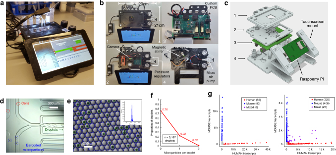
Droplet-based single-cell RNA-seq has emerged as a powerful technique for massively parallel cellular profiling. While this approach offers the exciting promise to deconvolute cellular heterogeneity in diseased tissues, the lack of cost-effective and user-friendly instrumentation has hindered widespread adoption of droplet microfluidic techniques. To address this, we developed a 3D-printed, low-cost droplet microfluidic control instrument and deploy it in a clinical environment to perform single-cell transcriptome profiling of disaggregated synovial tissue from five rheumatoid arthritis patients. We sequence 20,387 single cells revealing 13 transcriptomically distinct clusters. These encompass an unsupervised draft atlas of the autoimmune infiltrate that contribute to disease biology. Additionally, we identify previously uncharacterized fibroblast subpopulations and discern their spatial location within the synovium. We envision that this instrument will have broad utility in both research and clinical settings, enabling low-cost and routine application of microfluidic techniques.


Conflict of interest statement

The authors declare no competing financial interests.

Figures

Fig. 1
Fig. 1
Microfluidic control instrument design and validation. a Picture of the microfluidic control instrument performing a Drop-seq run. b Top down views of multi-tiered instrument. Levels 1–4 contain assorted components for instrument operation. c 3D rendering of the instrument with levels corresponding to those in b. Components in light gray are 3D printed. d Microscope image screen capture directly from the instrument. Cells and barcoded microparticles are visualized easily on the screen. e Microscope image of droplets output from the instrument. Droplets and microparticles are detected via image analysis software as blue circles and green circles respectively. Inset: droplet diameter distribution histogram. f Microparticle loading distribution into droplets as measured via automated image analysis is consistent with Poisson loading. g Species mixing experiments using mouse (3T3) and human (HEK293) cells at total cell concentrations of 75 cells/µl (left) and 300 cells/µl (right)
Fig. 2
Fig. 2
RA single-cell unsupervised clustering and analysis. a Sample workflow from operating room to sequencing. Preparation of single cells into droplets with barcoded microparticles is performed in about 2 h. b H&E stain of synovial tissue from patient tissue. The synovial lining is indicated by the black arrow. An example of vasculature is indicated by a red arrow. The blue arrow denotes a dense lymphocyte infiltrate. c Unsupervised graph-based clustering of single-cell RNA-seq, visualized using t-distributed stochastic neighbor embedding (tSNE). Each point represents a single cell (droplet barcode). d Graph-based clustering of single-cell RNA-seq, visualized using tSNE colored by patient sample. e Fraction of total cells present in each cluster across two patients (RA153 and RABP3) in replicate. f Fraction of total cells present in each cluster, for each patient. Color legend is as in e. g Bulk expression (‘in silico average’) comparisons across macrophages from each replicate for patients RA153 and RABP3. h Expression comparison across combined CD8+ T cell and macrophage populations from both replicates for patients RA153 and RABP3
Fig. 3
Fig. 3
Transcriptomic markers of gene expression for individual clusters. a Single cell expression heatmap displaying up to five transcriptomic markers for each cluster, based on differential expression testing. b Gene expression for canonical marker genes, overlaid on the tSNE visualization. A list of transcriptomic markers for each cluster is provided in Supplementary Data 1
Fig. 4
Fig. 4
Identification of synovial fibroblast subtypes. a Bulk expression (‘in silico average’) comparison of fibroblast populations (1 vs. 2a&b combined across all patients). Genes up-regulated in fibroblast population 1, based on differential expression analysis with Bonferroni-corrected p < 0.05, are indicated in red and genes expressed predominantly in fibroblast population 2 are indicated in teal. b Expression comparison across fibroblast sub-populations 2a and 2b. c CD55 (top) and THY1 (CD90) (bottom) expression across the global tSNE. d Pathway and gene set overdispersion analysis on the three fibroblast populations identified from unbiased clustering of single cell RNA-seq data from patient RABP3. Enrichment score corresponds to each cells’ first principle component loading from pathway analysis as computed in pagoda
Fig. 5
Fig. 5
Bulk RNA-seq and immunofluorescence of synovial fibroblast subtypes. a Cell surface expression of CD90, CD55 and podoplanin in synovial tissue. Synovial cells were gated on the CD45− PI− population (left panel) and analyzed for the proportion of CD90+ and CD55+ cells (middle panel), and relative podoplanin cell surface expression (right panel). b Bulk RNA-seq of flow sorted CD55+ and CD90+ patient samples RA195 and RABP3. Heatmap shows that markers of fibroblast populations identified in scRNA-seq data (Fig. 4a) are strongly differentially expressed in bulk. A list of transcriptomic markers for each sorted sample is provided in Supplementary Data 2. c We identified genes that were differentially expressed between CD55+ and CD90+ fibroblasts in both the bulk and single-cell datasets. The density of average log fold changes observed for these genes in the single-cell data is colored by upregulation in the bulk data, demonstrating agreement between the bulk and single-cell datasets. d CD90 and CD55 localization in RA synovial tissue. Paraffin-embedded synovial tissue was sectioned and assayed for target markers by immunofluorescent staining with antibodies for CD90 (green) or CD55 (green) and counterstained with DAPI (blue). Lymphocyte infiltrates are denoted by gray asterisks. Images were acquired at 20 × magnification. Scale bar is 100 µm

References

    1. Macosko EZ, et al. Highly parallel genome-wide expression profiling of individual cells using nanoliter droplets. Cell. 2015;161:1202–1214. doi: 10.1016/j.cell.2015.05.002. - DOI - PMC - PubMed
    1. Klein AM, et al. Droplet barcoding for single-cell transcriptomics applied to embryonic stem sells. Cell. 2015;161:1187–1201. doi: 10.1016/j.cell.2015.04.044. - DOI - PMC - PubMed
    1. Vitak SA, et al. Sequencing thousands of single-cell genomes with combinatorial indexing. Nat. Methods. 2017;14:302–8. doi: 10.1038/nmeth.4154. - DOI - PMC - PubMed
    1. Zheng GXY, et al. Massively parallel digital transcriptional profiling of single cells. Nat. Commun. 2017;8:1–12. doi: 10.1038/s41467-016-0009-6. - DOI - PMC - PubMed
    1. Shekhar K, et al. Comprehensive classification of retinal bipolar neurons by single-cell transcriptomics. Cell. 2016;166:1308–1323.e30. doi: 10.1016/j.cell.2016.07.054. - DOI - PMC - PubMed

Publication types

MeSH terms

LinkOut - more resources