

Zebrafish and medaka offer insights into the neurobehavioral correlates of vertebrate magnetoreception
- PMID: 29476093
- PMCID: PMC5824813
- DOI: 10.1038/s41467-018-03090-6
Zebrafish and medaka offer insights into the neurobehavioral correlates of vertebrate magnetoreception
Erratum in
-
Author Correction: Zebrafish and medaka offer insights into the neurobehavioral correlates of vertebrate magnetoreception.Nat Commun. 2018 Jul 17;9(1):2859. doi: 10.1038/s41467-018-05323-0. Nat Commun. 2018. PMID: 30018324 Free PMC article.
Abstract
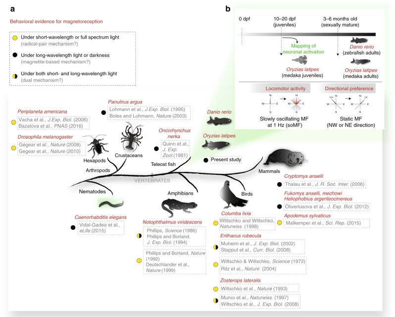
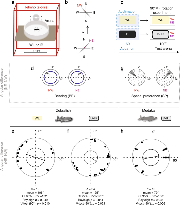
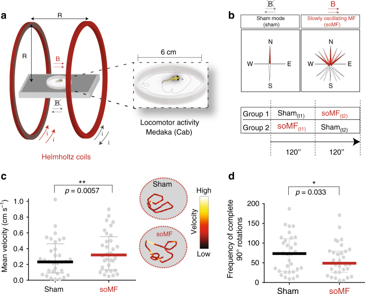
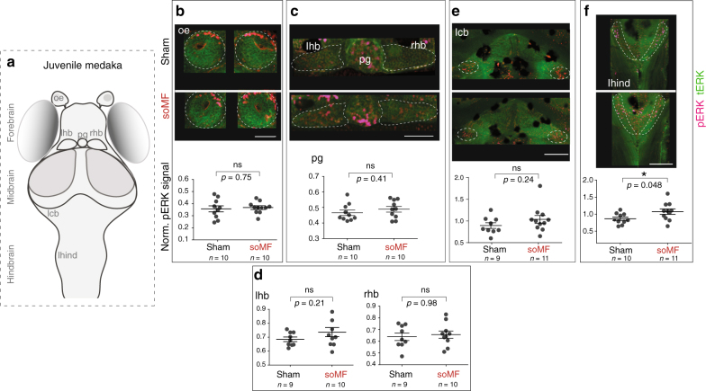
An impediment to a mechanistic understanding of how some species sense the geomagnetic field ("magnetoreception") is the lack of vertebrate genetic models that exhibit well-characterized magnetoreceptive behavior and are amenable to whole-brain analysis. We investigated the genetic model organisms zebrafish and medaka, whose young stages are transparent and optically accessible. In an unfamiliar environment, adult fish orient according to the directional change of a magnetic field even in darkness. To enable experiments also in juveniles, we applied slowly oscillating magnetic fields, aimed at generating conflicting sensory inputs during exploratory behavior. Medaka (but not zebrafish) increase their locomotor activity in this assay. Complementary brain activity mapping reveals neuronal activation in the lateral hindbrain during magnetic stimulation. These comparative data support magnetoreception in teleosts, provide evidence for a light-independent mechanism, and demonstrate the usefulness of zebrafish and medaka as genetic vertebrate models for studying the biophysical and neuronal mechanisms underlying magnetoreception.
Conflict of interest statement
The authors declare no competing financial interests.
Figures
Similar articles
-
Essential techniques for introducing medaka to a zebrafish laboratory--towards the combined use of medaka and zebrafish for further genetic dissection of the function of the vertebrate genome.Methods Mol Biol. 2011;770:211-41. doi: 10.1007/978-1-61779-210-6_8. Methods Mol Biol. 2011. PMID: 21805266
-
Zebrafish and medaka: model organisms for a comparative developmental approach of brain asymmetry.Philos Trans R Soc Lond B Biol Sci. 2009 Apr 12;364(1519):991-1003. doi: 10.1098/rstb.2008.0260. Philos Trans R Soc Lond B Biol Sci. 2009. PMID: 19064351 Free PMC article.
-
Small teleost fish provide new insights into human skeletal diseases.Methods Cell Biol. 2017;138:321-346. doi: 10.1016/bs.mcb.2016.09.001. Epub 2016 Oct 8. Methods Cell Biol. 2017. PMID: 28129851 Review.
-
Comparative aspects of gonadal sex differentiation in medaka: a conserved role of developing oocytes in sexual canalization.Sex Dev. 2009;3(2-3):99-107. doi: 10.1159/000223075. Epub 2009 Aug 10. Sex Dev. 2009. PMID: 19684455 Review.
-
Sensorimotor computation underlying phototaxis in zebrafish.Nat Commun. 2017 Sep 21;8(1):651. doi: 10.1038/s41467-017-00310-3. Nat Commun. 2017. PMID: 28935857 Free PMC article.
Cited by
-
Near absence of differential gene expression in the retina of rainbow trout after exposure to a magnetic pulse: implications for magnetoreception.Biol Lett. 2018 Jun;14(6):20180209. doi: 10.1098/rsbl.2018.0209. Biol Lett. 2018. PMID: 29875210 Free PMC article.
-
Proposed three-phenylalanine motif involved in magnetoreception signalling of an Actinopterygii protein expressed in mammalian cells.Open Biol. 2023 Nov;13(11):230019. doi: 10.1098/rsob.230019. Epub 2023 Nov 22. Open Biol. 2023. PMID: 37989224 Free PMC article.
-
Magnetic orientation in juvenile Atlantic herring (Clupea harengus) could involve cryptochrome 4 as a potential magnetoreceptor.J R Soc Interface. 2024 Jun;21(215):20240035. doi: 10.1098/rsif.2024.0035. Epub 2024 Jun 5. J R Soc Interface. 2024. PMID: 38835248 Free PMC article.
-
Indication of Electromagnetic Field Exposure via RBF-SVM Using Time-Series Features of Zebrafish Locomotion.Sensors (Basel). 2020 Aug 26;20(17):4818. doi: 10.3390/s20174818. Sensors (Basel). 2020. PMID: 32858993 Free PMC article.
-
Advanced visual components inspired by animal eyes.Nanophotonics. 2024 Mar 1;13(6):859-879. doi: 10.1515/nanoph-2024-0014. eCollection 2024 Mar. Nanophotonics. 2024. PMID: 39634370 Free PMC article. Review.
References
-
- Lohmann K, et al. Magnetic orientation of spiny lobsters in the ocean: experiments with undersea coil systems. J. Exp. Biol. 1995;198:2041–2048. - PubMed
Publication types
MeSH terms
LinkOut - more resources
Full Text Sources
Other Literature Sources
Molecular Biology Databases
