. 2018 Feb 23;8(1):3543.
doi: 10.1038/s41598-018-21801-3.

Sequential Membrane Rupture and Vesiculation during Plasmodium berghei Gametocyte Egress from the Red Blood Cell

Affiliations

Sequential Membrane Rupture and Vesiculation during Plasmodium berghei Gametocyte Egress from the Red Blood Cell

Maria Andreadaki et al. Sci Rep. .

Abstract

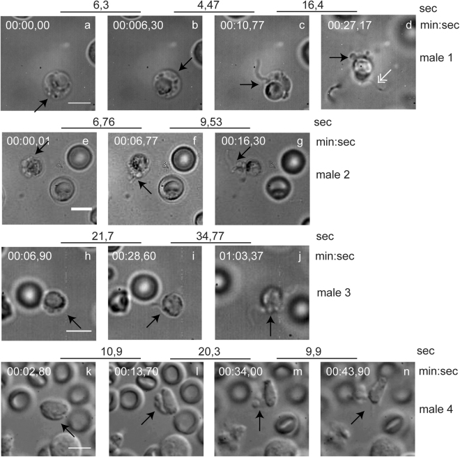
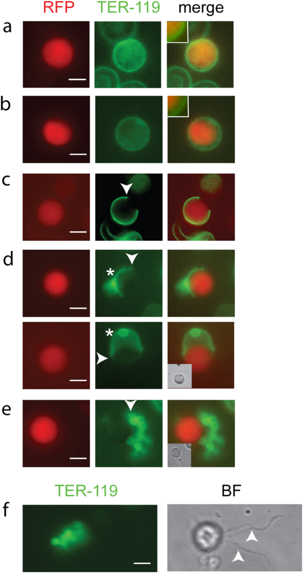
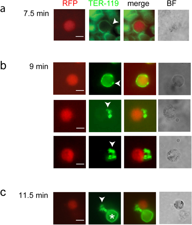
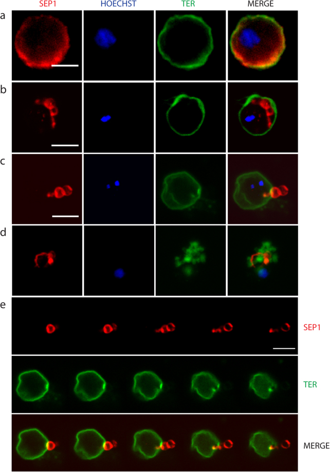
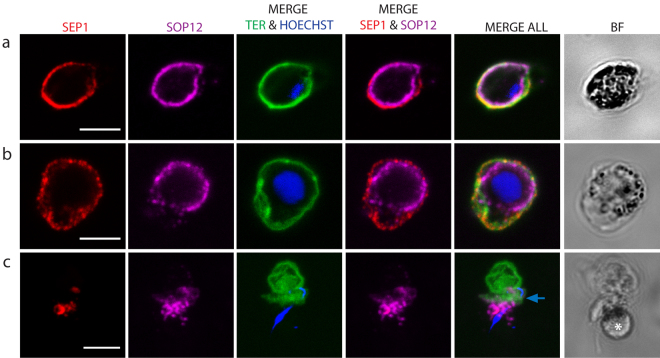
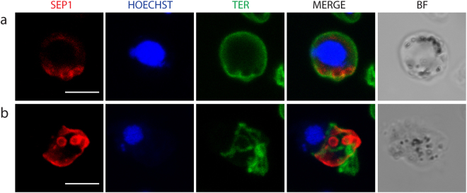
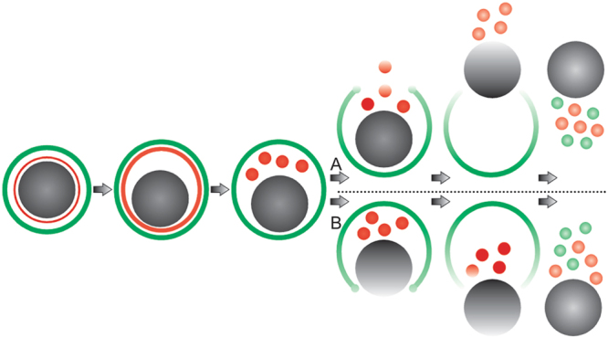
Malaria parasites alternate between intracellular and extracellular stages and successful egress from the host cell is crucial for continuation of the life cycle. We investigated egress of Plasmodium berghei gametocytes, an essential process taking place within a few minutes after uptake of a blood meal by the mosquito. Egress entails the rupture of two membranes surrounding the parasite: the parasitophorous vacuole membrane (PVM), and the red blood cell membrane (RBCM). High-speed video microscopy of 56 events revealed that egress in both genders comprises four well-defined phases, although each event is slightly different. The first phase is swelling of the host cell, followed by rupture and immediate vesiculation of the PVM. These vesicles are extruded through a single stabilized pore of the RBCM, and the latter is subsequently vesiculated releasing the free gametes. The time from PVM vesiculation to completion of egress varies between events. These observations were supported by immunofluorescence microscopy using antibodies against proteins of the RBCM and PVM. The combined results reveal dynamic re-organization of the membranes and the cortical cytoskeleton of the erythrocyte during egress.


Conflict of interest statement

The authors declare no competing interests.

Figures

Figure 1
Figure 1
Snapshots from four movies of female gamete egress. Representative snapshots chosen to illustrate the stages of egress. Upper broken lines represent time in sec between each frame of the movies in real time. The time of each frame from the start of the movies is indicated above the snapshots (min:sec). For technical reasons this does not correspond with the time from the start of the experiment (activation). (ad) Female 1. (a) An infected RBC in the initial phase of swelling. The parasite occupies most of the volume of the RBC. (b) The RBCM is more extended. (c) The PVM vesicles are extruded through a single pore. (d) Egress is complete. Vesiculated PVM and RBCM are localized in proximity to the gamete. (eh) Female 2. (e,f) The PVM is already vesiculated inside the RBCM. (g) The RBCM is extended and the gamete slides out of the host membrane. (h) Egress is complete with vesicles next to the gamete. (i–l) Female 3. (i) The RBCM is largely extended. (j) PVM is ruptured and vesiculated. (k) The gamete slides out of the extended RBCM. (l) egress is complete, the RBCM is vesiculated. (mp) Female 4. (m) RBC is swollen. (n) PVM vesicles are seen inside the RBCM towards the viewer. (o) Expulsion of PVM vesicles through a single pore. (p) Egress is complete. Black arrows indicate the area of interest in each snapshot, and white double arrows direction of gamete expulsion in (g) and (k). Scale bar, 5 μm. See also Supplementary Information Videos S1–S4.
Figure 2
Figure 2
Snapshots from movies of four male gametocytes undergoing egress and exflagellation. Representative snapshots chosen to illustrate the stages of egress. Upper broken lines represent time in sec between each frame of the movies in real time. The time of each frame from the start of the movies is indicated above the snapshots (min:sec). For technical reasons this does not correspond with the time from the start of the experiment (activation). (ad) Male 1. (a,b) The RBC is swollen with PVM vesicles visible. (c) A flagellum is exiting the RBCM through a single pore. (d) Egress is complete. Vesiculated PVM and RBCM are localized in proximity residual gametocyte and a free flagellum is also visible (white double arrow). (eg) Male 2. (e) The PVM is already vesiculated inside the RBCM. (f) A vesicle exits the RBCM through a single pore followed by a single flagellum. (g) Egress is complete with vesicles and a free flagellum next to the residual gametocyte. (hj) Male 3. (h) The RBCM is extended with PVM vesicles inside. (i) A single flagellum is exiting the RBCM through a single pore. (j) Egress is complete, with vesicles and one flagellum in proximity to the residual gametocytes. (kn) Male 4. (k) The RBCM is extended. (l) The PVM is vesiculated. (m) Vesicles of RBCM and PVM are seen close to the cell. (n) Exflagellation complete. Black arrows indicate the area of interest in each snapshot. Scale bar, 5 μm. See also Supplementary Information Videos S5–S9.
Figure 3
Figure 3
Live imaging reveals RBCM rupture. Samples were imaged without prior fixation. (ae) Female cells expressing RFP (red) and labeled with TER-119 (green). (a) A female gametocyte still enclosed in the intact RBCM. The membrane is adjacent to the parasites. (b) Swelling of the RBCM membrane is evident. Insets show the adjacent (a) and displaced RBCM (b) compared to the parasite. (c) RBCM opened at a single point. Arrow points to a slight thickening of the membrane at the opening. (d) Two cells in the process of egress. The arrows point to a slight thickening of the membrane at the opening. Top panel: The RBCM is opening and simultaneously forming vesicles (asterisks, out of focus), the most common RBCM rupture detected. Lower panel: The RBCM opens as an intact membrane. Compare Fig. 1, female 2 and 3 and Supplementary Information Videos S2, S3. (e) Rupture of RBCM completed, and RBCM vesicles remain next to the gamete (white arrow). White arrow heads in (ce) point to opened or vesiculated RBCM. (f) Egressed male which is exflagellating. RBCM vesicles remain in proximity to the residual gametocyte and two flagellar gametes are visible (BF image, white arrows). The cells are motile and thus slightly differently located between panels. Scale bar, 5 µm.
Figure 4
Figure 4
Outside-out vesiculation revealed with TER-119 label. (ac) Females (expressing RFP, red) were fixed at different time points after activation and the RBCM labeled with TER-119 (green). (a) 7.5 min after activation the RBCM has opened at a single site. (b) Three females having completed gametogenesis 9 min after activation. Labeled outside-out vesicles remain in proximity to the gametes. (c) Completed gametogenesis at 11.5 min. Labeled vesicles remain close to the gamete. An intact RBC is indicated with an asterisk. White arrows point to opened or vesiculated RBCM. Scale bar, 5 µm.
Figure 5
Figure 5
Labeling with SEP1 antibodies reveal PVM vesicles. Cells were labeled with the SEP1 antibody (red) and TER-119 (green) highlighting the PVM and RBCM, respectively. DNA is stained with Hoechst 33342 (blue). (ad) Are projections of sections from confocal imaging. (a) Non-activated cell. (b) An activated cell. PVM vesicles have formed inside the intact RBCM. (c) The RBCM is opened at a single pore and SEP1 labeled vesicles are in the process of exiting. A montage of the same cell is shown in e. (d) A cell where egress is completed. Vesicles of RBCM and PVM are intermingled. (e) Montage of single sections of the cell in (c). Scale bar, 5 µm.
Figure 6
Figure 6
Labeling of SOP12-EGFP gametocytes reveal swelling of PV. The PVM was labeled with the SEP1 antibody (red), the parasite membrane protein SOP12 is highlighted with a GFP antibody (magenta) and the RBCM labeled with TER-119 (green). DNA is stained with Hoechst 33342 (blue). (a) Non-activated gametocyte. SOP12 is localized adjacent to the PVM and RBCM. (b) Activated gametocyte. The RBCM and PVM are extended, indicating the swelling of the PV and shrinking of the RBC cytoplasm. SOP12 is delineating the gametocyte. (c) Exflagellating male. The SOP12 label is no longer clearly delineated suggesting that the protein is secreted. Blue arrow shows the RBC membrane vesicles and above them there is another intact RBC. White asterisk shows the parasite. Projections of confocal stacks through the middle of the cell in a and b, and through the whole cell in c. Scale bar, 5 µm.
Figure 7
Figure 7
Parasites lacking PPLP2 form PVM vesicles. Activated pplp2(-) gametocytes labeled with the SEP1 antibody (red) and TER-119 (green). DNA is stained with Hoechst 33342 (blue). The samples were treated with equinatoxin which destabilizes the RBCM. (a) SEP1 labeled vesicles are formed inside the intact RBCM. (b) SEP1 labeled vesicles close to a partially ruptured RBCM.
Figure 8
Figure 8
Electron microscopy analysis. (a) An activated male gametocyte. Inside the intact RBCM three vesicles are visible (V) probably derived from the PVM. No PVM surrounds the gametocyte. (b) Two activated gametocytes where the red blood cell cytoplasm has been completely digested. The RBCM is still intact. (c) Membrane spirals and whorls detected free in samples of activated gametocytes. (d,e) Scanning EM of activated gametocytes. (d) A cell is surrounded by a thin membrane collapsed on the grid. A convoluted empty membrane is located next to it. The rough surface suggests that the inner leaflet faces outwards. (e) An intact membrane sheet collapsed on a cell suggesting that the membrane was opened at one point and the opening then extended. The membrane is smooth contrasting to d. Ax, axoneme; N, nucleus; RBCM, red blood cell membrane; GAM, gamete.
Figure 9
Figure 9
New model for egress. The gametocyte (gray) is surrounded by the PVM (red) and RBCM (green). The first sign of egress is swelling of the two membranes. Next, rupture and vesiculation of the PVM take place followed by the opening of a single stabilized pore in the RBCM. Two alternative positions of the pore were seen. The most common case was localization of the pore towards the PVM vesicles (A), which was seen in most females and all males. In more rare cases, and only detected in females, the pore was sited towards the parasite (B). Curling of the pore was suggested by the thickening of the rim detected in some events. The last step is the vesiculation of the RBCM resulting in a free female gamete or male residual cell.

Similar articles

Cited by

References

    1. Deligianni E, et al. A perforin-like protein mediates disruption of the erythrocyte membrane during egress of Plasmodium berghei male gametocytes. Cellular microbiology. 2013;15:1438–1455. doi: 10.1111/cmi.12131. - DOI - PubMed
    1. Sologub L, et al. Malaria proteases mediate inside-out egress of gametocytes from red blood cells following parasite transmission to the mosquito. Cellular microbiology. 2011;13:897–912. doi: 10.1111/j.1462-5822.2011.01588.x. - DOI - PubMed
    1. Spielmann T, Montagna GN, Hecht L, Matuschewski K. Molecular make-up of the Plasmodium parasitophorous vacuolar membrane. International journal of medical microbiology: IJMM. 2012;302:179–186. doi: 10.1016/j.ijmm.2012.07.011. - DOI - PubMed
    1. Matthews K, et al. The Plasmodium translocon of exported proteins (PTEX) component thioredoxin-2 is important for maintaining normal blood-stage growth. Molecular microbiology. 2013;89:1167–1186. doi: 10.1111/mmi.12334. - DOI - PubMed
    1. Baker DA, Daramola O, McCrossan MV, Harmer J, Targett GA. Subcellular localization of Pfs16, a Plasmodium falciparum gametocyte antigen. Parasitology. 1994;108(Pt 2):129–137. doi: 10.1017/S0031182000068219. - DOI - PubMed

Publication types

MeSH terms

Substances