. 2018 Aug;28(8):3362-3371.
doi: 10.1007/s00330-017-5265-2. Epub 2018 Feb 23.

How reliable are ADC measurements? A phantom and clinical study of cervical lymph nodes

Affiliations

How reliable are ADC measurements? A phantom and clinical study of cervical lymph nodes

Bastien Moreau et al. Eur Radiol. 2018 Aug.

Abstract

Objective: To assess the reliability of ADC measurements in vitro and in cervical lymph nodes of healthy volunteers.

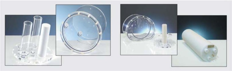
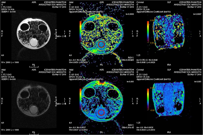
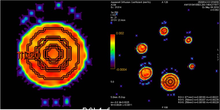
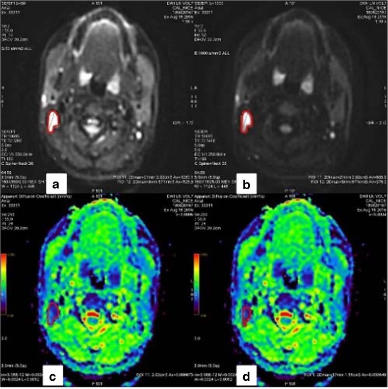
Methods: We used a GE 1.5 T MRI scanner and a first ice-water phantom according to recommendations released by the Quantitative Imaging Biomarker Alliance (QIBA) for assessing ADC against reference values. We analysed the target size effect by using a second phantom made of six inserted spheres with diameters ranging from 10 to 37 mm. Thirteen healthy volunteers were also scanned to assess the inter- and intra-observer reproducibility of volumetric ADC measurements of cervical lymph nodes.

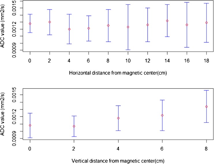
Results: On the ice-water phantom, the error in ADC measurements was less than 4.3 %. The spatial bias due to the non-linearity of gradient fields was found to be 24 % at 8 cm from the isocentre. ADC measure reliability decreased when addressing small targets due to partial volume effects (up to 12.8 %). The mean ADC value of cervical lymph nodes was 0.87.10-3 ± 0.12.10-3 mm2/s with a good intra-observer reliability. Inter-observer reproducibility featured a bias of -5.5 % due to segmentation issues.

Conclusion: ADC is a potentially important imaging biomarker in oncology; however, variability issues preclude its broader adoption. Reliable use of ADC requires technical advances and systematic quality control.

Key points: • ADC is a promising quantitative imaging biomarker. • ADC has a fair inter-reader variability and good intra-reader variability. • Partial volume effect, post-processing software and non-linearity of scanners are limiting factors. • No threshold values for detecting cervical lymph node malignancy can be drawn.

Keywords: Biomarkers; Diffusion; Lymph; Magnetic resonance imaging; Quantitative evaluation.


Conflict of interest statement

Guarantor

The scientific guarantor of this publication is Dr. Bastien Moreau, PhD.

Conflict of interest

Hubert Beaumont, as co-author of this manuscript, declares relationships with the following companies: Median Technologies.

All other authors of this manuscript declare no relationships with any companies whose products or services may be related to the subject matter of the article.

Statistics and biometry

One of the authors has significant statistical expertise.

Informed consent

Written informed consent was obtained from all subjects (patients) in this study.

Ethical approval

Institutional Review Board approval was obtained.

Methodology

• prospective

• experimental

• performed at one institution

Figures

Fig. 1
Fig. 1
Phantoms used in the study. Left ICEWATER phantom filled with 0° C water (DIN 6858-1, PTW, Freiburg, Germany). Right SPHERE phantom at room temperature featuring spheres of various sizes between 10- and 37-mm diameters (NEMA NU2-2012, PTW, Freiburg, Germany)
Fig. 2
Fig. 2
Measurements on the ICEWATER phantom. Imaging of ICEWATER phantom at b0-b100 (top) and b0-b800 (bottom). From left to right: Diffusion mapping, axial view of ADC mapping and coronal view of ADC mapping. Red circular regions of interest are set at the centre of the ice water cylinder
Fig. 3
Fig. 3
Measurements on the SPHERE phantom. Left Spherical VOIs of decreasing sizes centered on the largest sphere. Right Spherical volumes of interest (VOIs) centered on sphere of various sizes. VOIs diameters are set to 80 % of physical sphere’s diameters
Fig. 4
Fig. 4
Measurements of cervical lymphnodes. Imaging of a healthy volunteer’s cervical lymph node. (a) Diffusion mapping at b = 50. (b) mapping at b = 1000 on which the volume of interest (VOI) is contoured before being exported to other series. (c) and (d) Mapping of the apparent diffusion coefficient. In red, the VOI is determined by operator 1 (a, b and c) and by operator 2 (d)
Fig. 5
Fig. 5
Spatial correlation of ADC. Top view apparent diffusion coefficient (ADC) changes according to the horizontal distance from magnetic centre. Horizontal axis distance in cm. Vertical axis ADC value in mm2/s. Bottom view ADC changes according to the vertical distance from magnetic center. Horizontal axis Distance in cm. Vertical axis ADC value in mm2/s
Fig. 6
Fig. 6
Example of inter-observer discordance in terms of volume and apparent diffusion coefficient (ADC). Example of inter-observer discordance in terms of volume and ADC on a level II lymph node. Top row First reader’s measurements. Bottom row Second reader’s measurements. Left b1000 diffusion maps where volumes of interest (VOIs) are drawn. Right corresponding ADC maps. Note: The heterogeneity of the node’s environment featuring areas of high ADC values (green and red in the right images) without clear correspondence on the diffusion image

References

    1. Kessler LG, Barnhart HX, Buckler AJ, et al. The emerging science of quantitative imaging biomarkers terminology and definitions for scientific studies and regulatory submissions. Stat Methods Med Res. 2015;24:9–26. doi: 10.1177/0962280214537333. - DOI - PubMed
    1. Sullivan DC, Obuchowski NA, Kessler LG, et al. Metrology standards for quantitative imaging biomarkers. Radiology. 2015;277:813–825. doi: 10.1148/radiol.2015142202. - DOI - PMC - PubMed
    1. Thomassin-Naggara I, Dechoux S, Bonneau C, et al. How to differentiate benign from malignant myometrial tumours using MR imaging. Eur Radiol. 2013;23:2306–2314. doi: 10.1007/s00330-013-2819-9. - DOI - PubMed
    1. Hambrock T, Somford DM, Huisman HJ, et al. Relationship between apparent diffusion coefficients at 3.0-T MR imaging and Gleason grade in peripheral zone prostate cancer. Radiology. 2011;259:453–461. doi: 10.1148/radiol.11091409. - DOI - PubMed
    1. Higano S, Yun X, Kumabe T, et al. Malignant astrocytic tumors clinical importance of apparent diffusion coefficient in prediction of grade and prognosis. Radiology. 2006;241:839–846. doi: 10.1148/radiol.2413051276. - DOI - PubMed

Publication types