Review
. 2018 Apr;9(2):199-209.
doi: 10.1007/s13244-017-0590-1. Epub 2018 Feb 23.

Hyperechoic breast images: all that glitters is not gold!

Affiliations
Review

Hyperechoic breast images: all that glitters is not gold!

Gabrielle Journo et al. Insights Imaging. 2018 Apr.

Abstract

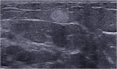
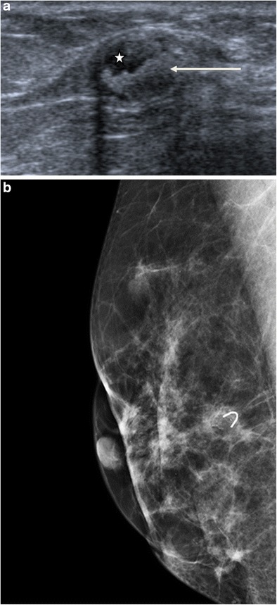
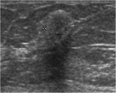
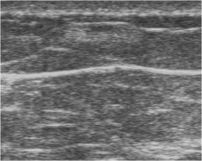
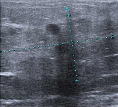
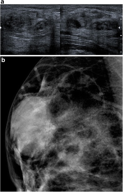
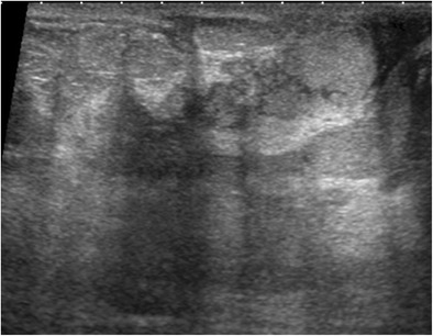
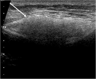
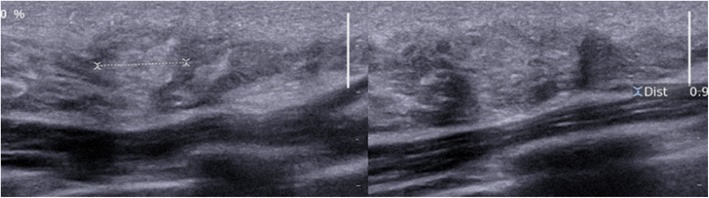
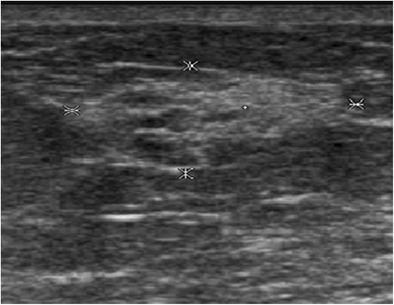
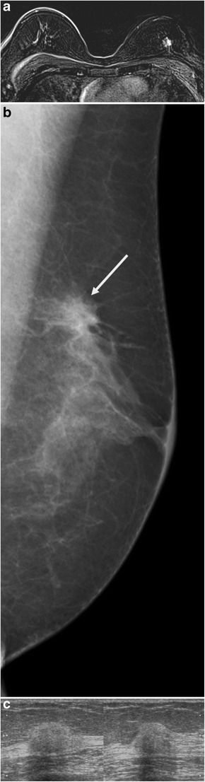
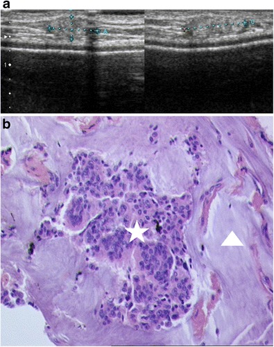
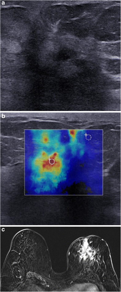
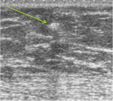
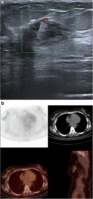
Hyperechogenicity is a sign classically reported to be in favour of a benign lesion and can be observed in many types of benign breast lesions such as hamartoma, lipoma, angiolipoma, haemangioma, haematoma, fat necrosis, fibrosis and galactocele, among others. However, some rare malignant breast lesions can also present a hyperechoic appearance. Most of these hyperechoic malignant lesions present other characteristics that are more typically suggestive of malignancy such as posterior shadowing, a more vertical axis or irregular margins that help to guide the diagnosis. Post magnetic resonance imaging, second-look ultrasound may visualise hyperechoic malignant lesions that would not have been identified at first sight and radiologists must know how to recognise these lesions.

Teaching points: • Some rare malignant breast lesions can present a hyperechoic appearance. • Malignant lesions present other characteristics that are suggestive of malignancy. • An echogenic mass with fat density on mammography does not require biopsy.

Keywords: Breast cancer; Breast ultrasound; Histology; Hyperechogenicity; Malignancy.


Figures

Fig. 1
Fig. 1
Spindle cell lipoma. Transverse ultrasound scan showing a homogeneous, hyperechoic, round mass with well circumscribed margins
Fig. 2
Fig. 2
Angiolipoma. Transverse ultrasound scan of the right breast showing a homogeneous, hyperechoic, oval-shaped subcutaneous mass with circumscribed margins
Fig. 3
Fig. 3
Fat necrosis. a Ultrasound scan showing a complex mass composed of an echo-free fat component (asterisk) and an echogenic fibrous component (arrow). b Mammography, oblique view, showing a radiolucent formation projected over a clip
Fig. 4
Fig. 4
Myofibroblastoma. Ultrasound scan showing a slightly heterogeneous, hyperechoic and attenuating rounded mass
Fig. 5
Fig. 5
Haemangioma in a 72-year-old woman, presenting with a slowly growing, superficial breast opacity. Ultrasound: oval-shaped, hyperechoic mass of the subcutaneous fat, parallel to the skin. Surgical resection in order to eliminate angiosarcoma: confirmation of the benign nature of the tumour
Fig. 6
Fig. 6
Haematoma. Patient followed for right breast cancer treated in 1998, referred for opinion concerning a mammographic opacity of the right breast not present on the previous mammogram. Ultrasound revealed a mixed hyperechoic and hypoechoic, partially attenuating zone. On clinical interview, the patient reported a motor vehicle accident with ecchymosis of the breast in contact with the safety belt. Ultrasound-guided microbiopsy demonstrated fat necrosis and no suspicious image. Regression of the image on regular follow-up examinations
Fig. 7
Fig. 7
Hamartoma. a Ultrasound showing an oval-shaped mass with mixed echogenicity, globally isoechoic, with an edge effect artefact. b Mammography showing a well-circumscribed, dense, oval-shaped mass, with a slightly heterogeneous appearance related to its mixed content (fat and stroma)
Fig. 8
Fig. 8
Galactocele. a Ultrasound scan showing duct dilatation with homogeneous hyperechoic content, with no blood supply demonstrated by colour Doppler, in a schizophrenic patient with long-term neuroleptic treatment. b Digital breast tomosynthesis in the same patient showing a fat density galactocele (arrow)
Fig. 9
Fig. 9
PASH. a Ultrasound scan showing a hyperechoic, slightly heterogeneous and attenuating oval-shaped mass. b Microscopy (HES ×20): histological section showing fibrosis with pseudoangiomatous stromal hyperplasia (arrow showing a channel lined by flat endothelium-like cells)
Fig. 10
Fig. 10
Abscess. A 42-year-old woman presenting with enlargement of the left breast for 1 month with pain and erythema related to mastitis of the left breast. Ultrasound scan of the left breast in the same patient revealing a mixed echogenicity, globally hyperechoic abscess
Fig. 11
Fig. 11
Siliconoma. Periprosthetic silicone (arrow showing a hyperechoic mass with attenuation of the ultrasound beam)
Fig. 12
Fig. 12
Complex sclerosing lesions. Ultrasound in a young woman with a palpable mass, revealing a hyperechoic zone with irregular margins and acoustic shadow. Microbiopsy demonstrating benign complex sclerosing lesions comprising duct structures and hyperplastic epithelium with no suspicious atypia, together with several pseudopapillary structures, and locally inflammatory periductal fibrosis
Fig. 13
Fig. 13
Adenosis. Internal swelling of the right breast with a diameter of 15 mm in a 45-year-old woman, corresponding to a poorly demarcated mammographic opacity associated with punctate microcalcifications, evolving very slowly over a period of 4 years. Ultrasound: zone of mixed echogenicity, predominantly hyperechoic, with a long axis parallel to the skin, not modifying the ultrasound beam
Fig. 14
Fig. 14
Invasive ductal carcinoma. Annual surveillance in a patient at very high risk already treated for left breast cancer. a MRI, gadolinium-enhanced T1 subtraction sequence, demonstrating an irregular, poorly circumscribed mass at the junction of the upper quadrants of the left breast. The gadolinium-enhanced examination shows initial 90% enhancement with washout at the late phase. b Mammography guided by MRI in the same patient, oblique view: demonstration of a non-calcified mass (arrows), with architectural distortion at the junction of the upper quadrants of the left breast. c MRI-guided ultrasound of the left breast in the same patient. At the junction of the upper quadrants, at the 12.30 position, 4 cm from the nipple, presence of a hyperechoic nodule with microlobular margins, with posterior attenuation classified as high ACR4. Microbiopsy demonstrated poorly differentiated invasive ductal carcinoma (grade II, low MI, HR+, HER2-)
Fig. 15
Fig. 15
Mucinous carcinoma. Patient with a local subcutaneous recurrence in the upper inner quadrant, 10 years after mastectomy with secondary prosthetic reconstruction. a Ultrasound: hyperechoic mass of the subcutaneous tissue, with slightly microlobular margins. b Histological examination of an ultrasound-guided microbiopsy (HES ×20): clumps of tumour cells (asterisk) surrounded by zones of mucin (arrowhead)
Fig. 16
Fig. 16
Invasive lobular carcinoma. a Mammography, AP view, showing a mass with irregular margins in the right upper inner quadrant (arrows). b Ultrasound showing an irregular, poorly circumscribed, vertically oriented, hyperechoic and partially attenuating lesion
Fig. 17
Fig. 17
Angiosarcoma. A 76-year-old woman, treated conservatively for left breast cancer in 1993. Palpation of an upper quadrant breast swelling. a Ultrasound: heterogeneous, hyperechoic mass, with a hypoechoic centre. b Elastography, zones of hardness (red) in the periphery of the mass. c MRI, axial gadolinium-enhanced T1 subtraction sequence: spiculated mass with heterogeneous intense contrast enhancement. Histological examination revealed a high-grade moderately to poorly differentiated angiosarcoma
Fig. 18
Fig. 18
Metastasis. Ultrasound showing a small hyperechoic, attenuating mass (arrow) in a patient followed for malignant melanoma. Cytological examination after ultrasound-guided fine-needle aspiration: very numerous spindle-shaped, pigmented, round malignant cells, corresponding to a metastasis from known malignant melanoma in this patient
Fig. 19
Fig. 19
Lymphoma. a Ultrasound of the right breast showing a poorly demarcated, mixed echogenicity mass, with blood flow on Doppler ultrasound. b PET-CT scan in this patient shows a hypermetabolic lesion in the right breast. Microbiopsy revealed diffuse large B-cell lymphoma

References

    1. American College of Radiology . Illustrated breast imaging reporting and data system (BI-RADS) 3. Reston: American College of Radiology; 1998.
    1. Skaane P, Engedal K. Analysis of sonographic features in the differentiation of fibroadenoma and invasive ductal carcinoma. AJR Am J Roentgenol. 1998;170:109–104. doi: 10.2214/ajr.170.1.9423610. - DOI - PubMed
    1. Linda A, Zuiani C, Lorenzon M, Furlan A, Londero V, Machin P, et al. The wide spectrum of hyperechoic lesions of the breast. Clin Radiol. 2011;66:559–565. doi: 10.1016/j.crad.2010.12.013. - DOI - PubMed
    1. Stavros AT, Thickman D, Rapp CL, Dennis MA, Parker SH, Sisney GA. Solid breast nodules: use of sonography to distinguish between benign and malignant lesions. Radiology. 1995;196:123–134. doi: 10.1148/radiology.196.1.7784555. - DOI - PubMed
    1. Mendelson EB, Böhm-Vélez M, Berg WA, et al. ACR BI-RADS® ultrasound. In: ACR BI-RADS® atlas, breast imaging reporting and data system. Reston: American College of Radiology; 2013.

LinkOut - more resources