. 2018 Apr;285(8):1477-1490.
doi: 10.1111/febs.14421. Epub 2018 Mar 8.

Crystal structures of sampatrilat and sampatrilat-Asp in complex with human ACE - a molecular basis for domain selectivity

Affiliations

Crystal structures of sampatrilat and sampatrilat-Asp in complex with human ACE - a molecular basis for domain selectivity

Gyles E Cozier et al. FEBS J. 2018 Apr.

Abstract

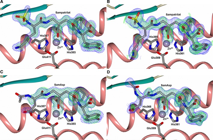
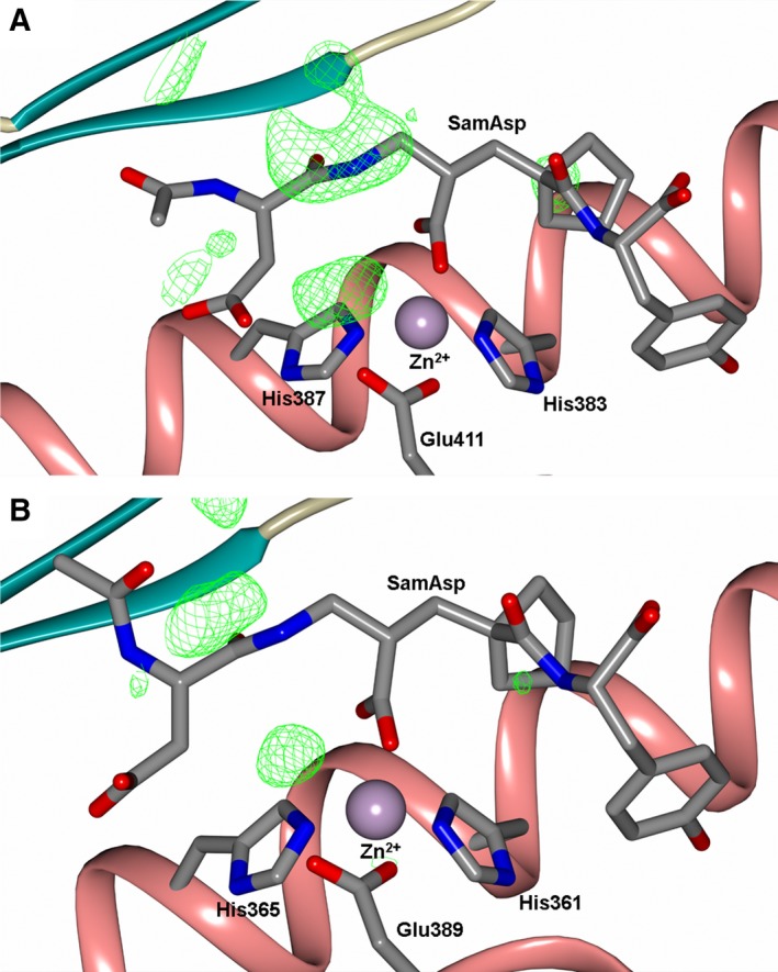
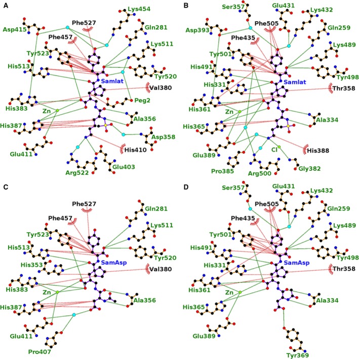
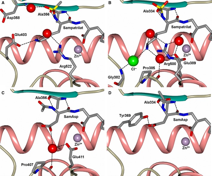
Angiotensin-1-converting enzyme (ACE) is a zinc metallopeptidase that consists of two homologous catalytic domains (known as nACE and cACE) with different substrate specificities. Based on kinetic studies it was previously reported that sampatrilat, a tight-binding inhibitor of ACE, Ki = 13.8 nm and 171.9 nm for cACE and nACE respectively [Sharma et al., Journal of Chemical Information and Modeling (2016), 56, 2486-2494], was 12.4-fold more selective for cACE. In addition, samAsp, in which an aspartate group replaces the sampatrilat lysine, was found to be a nonspecific and lower micromolar affinity inhibitor. Here, we report a detailed three-dimensional structural analysis of sampatrilat and samAsp binding to ACE using high-resolution crystal structures elucidated by X-ray crystallography, which provides a molecular basis for differences in inhibitor affinity and selectivity for nACE and cACE. The structures show that the specificity of sampatrilat can be explained by increased hydrophobic interactions and a H-bond from Glu403 of cACE with the lysine side chain of sampatrilat that are not observed in nACE. In addition, the structures clearly show a significantly greater number of hydrophilic and hydrophobic interactions with sampatrilat compared to samAsp in both cACE and nACE consistent with the difference in affinities. Our findings provide new experimental insights into ligand binding at the active site pockets that are important for the design of highly specific domain selective inhibitors of ACE.

Database: The atomic coordinates and structure factors for N- and C-domains of ACE bound to sampatrilat and sampatrilat-Asp complexes (6F9V, 6F9R, 6F9T and 6F9U respectively) have been deposited in the Protein Data Bank, Research Collaboratory for Structural Bioinformatics, Rutgers University, New Brunswick, NJ (http://www.rcsb.org/).

Keywords: angiotensin-1-converting enzyme; crystallography; domain specificity; enzyme kinetics; enzyme structure; metalloprotease; sampatrilat.


Figures

Figure 1
Figure 1
Schematic representation of inhibitors bound to the ACE domains overlayed with the final 2mFo‐DFc (blue, contoured at 1σ level) electron density map and the mFo‐DFc (green, contoured at 3σ level) electron density omit map for (A) Sampatrilat‐cACE, (B) Sampatrilat‐nACE, (C) SamAsp‐cACE and (D) SamAsp‐nACE complexes. The zinc ion is shown as a lilac sphere with the coordinating side chains shown as sticks. Alpha‐helices and β‐strands are shown in rose and dark cyan respectively.
Figure 2
Figure 2
Schematic representation of (A) SamAsp‐cACE‐ and (B) SamAsp‐nACE‐binding sites overlayed with the final mFo‐DFc (green, contoured at 3σ level) electron density difference map highlighting the electron density that is maybe from an alternate conformation of the samAsp P1/P2 groups. The zinc ion is shown as a lilac sphere with the coordinating side chains shown as sticks. Alpha‐helices and β‐strands are shown in rose and dark cyan respectively.
Figure 3
Figure 3
Residues 20–24 of nACE from the sampatrilat complex structure to highlight the second conformation of part of the nACE hinge region observed. (A) Both conformations, (B) Predominant conformation and (C) Alternate conformation overlayed with the 2mFo‐DFc (blue, contoured at 1σ level) and mFo‐DFc (green, contoured at 3σ level) electron density omit maps.
Figure 4
Figure 4
Two views of a schematic representation of the nACE protein chain from the sampatrilat complex structure showing the two conformations of part of the hinge region (residues 4–43 and 57–99) in fat worm style (predominant and alternate conformations shown in orange and blue respectively). Alpha‐helices are shown as tubes, sampatrilat as sticks and the zinc ion as a lilac sphere.
Figure 5
Figure 5
Modification of Cys496 from the tACE‐sampatrilat complex structure. (A,B) and (C) show the 2mFo‐DFc (blue, contoured at 1σ level) and mFo‐DFc (green, contoured at 3σ level) electron density omit maps generated from the first 5700, 2000 and 1500 images respectively. (D) The modelled oxidized cysteine residue Cso (S‐Hydroxycysteine) overlayed with the final 2mFo‐DFc (blue, contoured at 1σ level) electron density map. There is no density visible around this residue in the mFo‐DFc difference electron density map contoured at 3σ level indicating the oxidized cysteine fits the electron density map.
Figure 6
Figure 6
Ligplot representation of the binding site interactions of (A) sampatrilat‐cACE, (B) sampatrilat‐nACE, (C) samAsp‐cACE and (D) samAsp‐nACE. H‐bond/electrostatic interactions are shown in green, hydrophobic interactions in red and water molecules as cyan spheres. Residues solely involved in hydrophobic interactions are represented by red, semicircular symbols.
Figure 7
Figure 7
Close up views of (A) sampatrilat‐cACE, (B) sampatrilat‐nACE, (C) samAsp‐cACE and (D) samAsp‐nACE binding sites showing the H‐bond/electrostatic interactions in the S1 and S2 subsites. The protein chain is shown as a cartoon with α‐helices and β‐strands in rose and dark cyan, respectively, and water molecules are depicted as red spheres.
Figure 8
Figure 8
(A) Structure of RXPA380. (B) Ligplot representation of the binding site interactions of RXPA380 in cACE. H‐bond/electrostatic interactions are shown in green, hydrophobic interactions in red and water molecules as cyan spheres.

References

    1. Acharya KR, Sturrock ED, Riordan JF & Ehlers MR (2003) Ace revisited: a new target for structure‐based drug design. Nat Rev Drug Discov 2, 891–902. - PMC - PubMed
    1. Skeggs LT Jr, Kahn JR & Shumway NP (1956) The preparation and function of the hypertensin‐converting enzyme. J Exp Med 103, 295–299. - PMC - PubMed
    1. Yang HY, Erdos EG & Levin Y (1970) A dipeptidyl carboxypeptidase that converts angiotensin I and inactivates bradykinin. Biochim Biophys Acta 214, 374–376. - PubMed
    1. Araujo MC, Melo RL, Cesari MH, Juliano MA, Juliano L & Carmona AK (2000) Peptidase specificity characterization of C‐ and N‐terminal catalytic sites of angiotensin I‐converting enzyme. Biochemistry 39, 8519–8525. - PubMed
    1. Rousseau A, Michaud A, Chauvet MT, Lenfant M & Corvol P (1995) The hemoregulatory peptide N‐acetyl‐Ser‐Asp‐Lys‐Pro is a natural and specific substrate of the N‐terminal active site of human angiotensin‐converting enzyme. J Biol Chem 270, 3656–3661. - PubMed

Publication types

MeSH terms