. 2018 May;188(5):1149-1160.
doi: 10.1016/j.ajpath.2018.01.021. Epub 2018 Feb 21.

A Conserved Distal Lung Regenerative Pathway in Acute Lung Injury

Affiliations

A Conserved Distal Lung Regenerative Pathway in Acute Lung Injury

Martin S Taylor et al. Am J Pathol. 2018 May.

Abstract

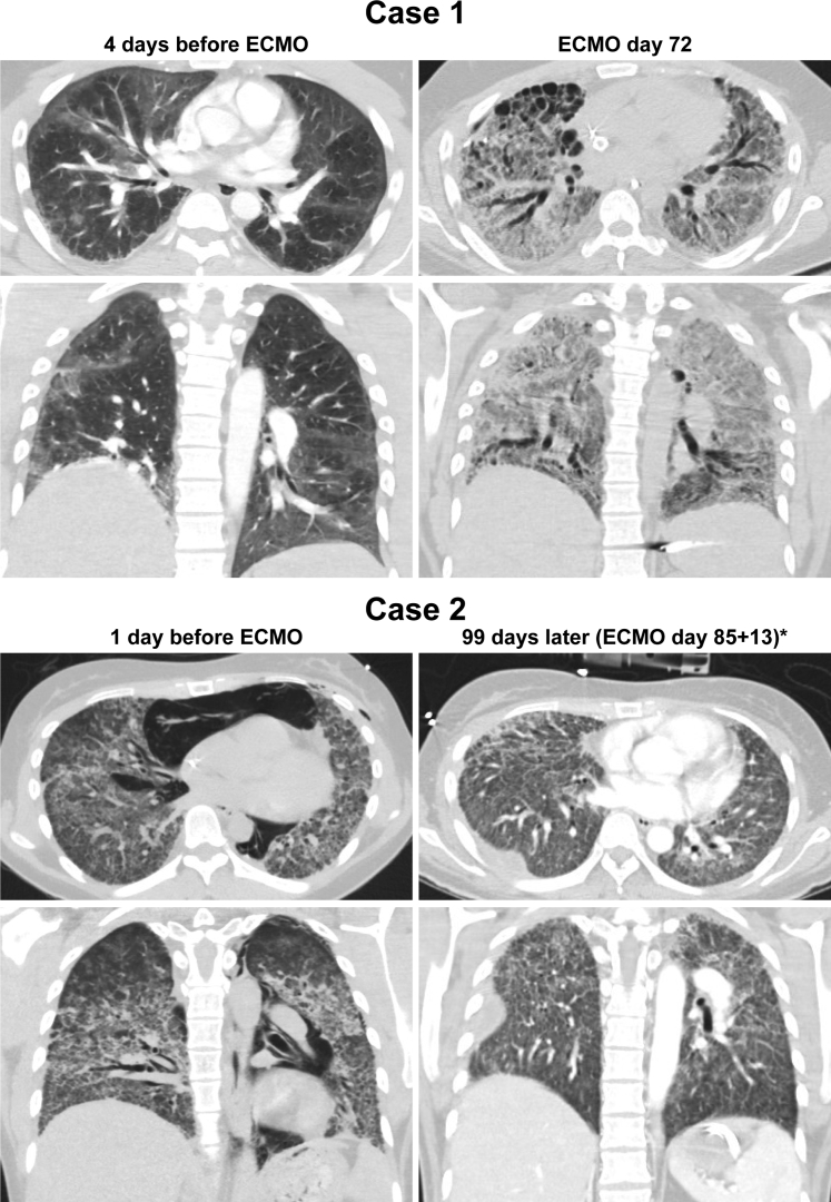
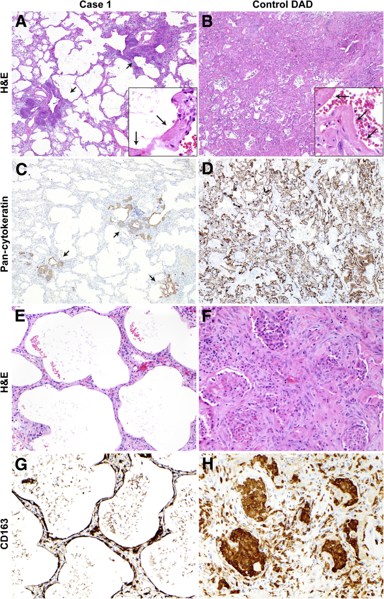
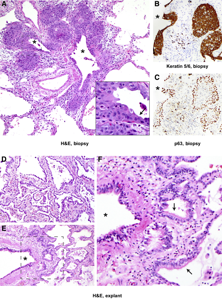
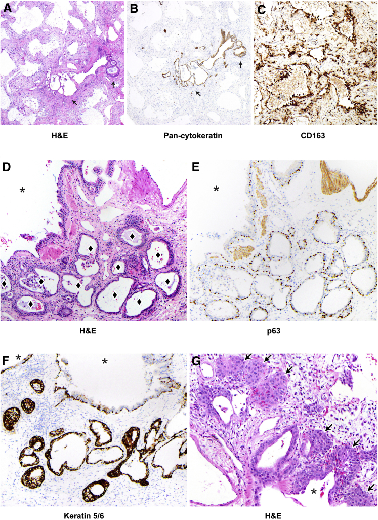
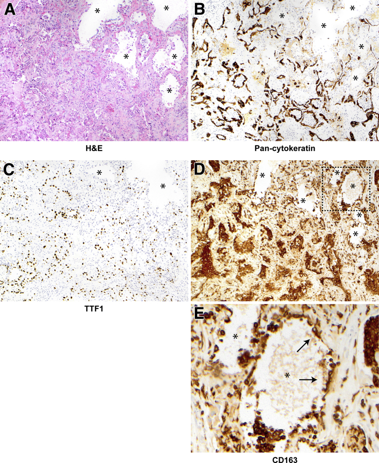
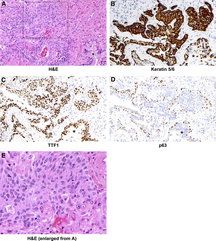
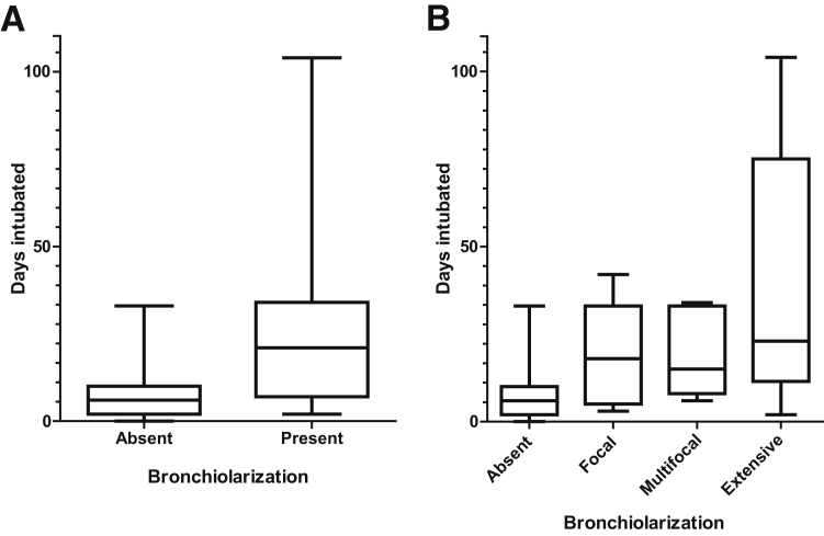
Improved tools have led to a burgeoning understanding of lung regeneration in mice, but it is not yet known how these insights may be relevant to acute lung injury in humans. We report in detail two cases of fulminant idiopathic acute lung injury requiring extracorporeal membrane oxygenation in previously healthy young adults with acute respiratory distress syndrome, one of whom required lung transplantation. Biopsy specimens showed diffuse alveolar injury with a striking paucity of alveolar epithelial regeneration, rare hyaline membranes, and diffuse contiguous airspace lining by macrophages. This novel constellation was termed diffuse alveolar injury with delayed epithelization. In addition, mirroring data from murine models of lung injury/regeneration, peribronchiolar basaloid pods (previously described as squamous metaplasia) and ciliated bronchiolarization were identified in these patients and in 39% of 57 historical cases with diffuse alveolar damage. These findings demonstrate a common and clinically relevant human disease correlate for murine models of severe acute lung injury. Evidence suggests that peribronchiolar basaloid pods and bronchiolarization are related spatially and temporally and likely represent overlapping sequential stages of the response to severe distal airway injury.


Figures

Figure 1
Figure 1
Computed tomographic scans. The lungs in case 1 before extracorporeal membrane oxygenation (ECMO) show patchy bilateral ground-glass opacities and plate-like atelectasis. At ECMO day 72 and before orthotopic lung transplantation, there is diffuse interstitial fibrosis, traction bronchiectasis, and subpleural honeycomb cystic change. The lungs in case 2 before ECMO demonstrate pneumomediastinum and bilateral interstitial opacities with centrilobular accentuation. Ninety-eight days after ECMO initiation (asterisk, after 85 days of ECMO and 13 days decannulated), there is bilateral interstitial fibrosis and a loculated right effusion.
Figure 2
Figure 2
Alveolar epithelialization in diffuse alveolar injury with delayed epithelization case 1 versus control diffuse alveolar damage (DAD), matched in timing relative to the onset of clinical acute respiratory distress syndrome. A: Lung sections stained and examined at low magnification demonstrate diffuse alveolar injury and peribronchiolar basaloid pods (short arrows). Hyaline membranes are rare (long arrows, inset), and the lung interstitium is mildly thickened. B: Control case of DAD in the proliferative phase shows diffuse hyaline membranes (arrows, inset) and thickened lung interstitium. C: The absence of TTF1+/cytokeratin+ alveolar epithelium in case 1 is highlighted by the lack of cytokeratin cellular immunostaining; peribronchiolar basaloid pods are cytokeratin positive (arrows). D: In contrast, there is diffuse cytokeratin immunostaining of alveolar lining cells and hyaline membranes in the control. E: Denuded alveoli in case 1 show a lack of alveolar epithelium, with no hyaline membranes in this region. F and H: In control DAD, there are abundant CD163+ intra-alveolar and interstitial macrophages. G: CD163 immunohistochemistry demonstrates macrophages contiguously lining alveoli and in the interstitium. Original magnification: ×40 (AD); ×200 (EH); ×400 (insets). H&E, hematoxylin and eosin.
Figure 3
Figure 3
Peribronchiolar basaloid pods (previously referred to as squamous metaplasia in the pathology literature5, 14, 15) and bronchiolarization in biopsy and explants, case 1. A: Proliferating basaloid/squamoid cells are adjacent to a terminal bronchiole and are focally contiguous with ciliated bronchiolar epithelium (arrow indicates ciliated bronchiolar cells; asterisks, bronchiolar lumens; inset, higher magnification of boxed area). B and C: Pod cells are immunopositive for TTF1 (not shown) and keratin 5/6 (B), and basally positive for p63 (C). Asterisks indicate bronchiolar lumens. D: Explanted lungs in case 1 after 101 days on extracorporeal membrane oxygenation demonstrate robust alveolar reepithelialization and fibrosis. E and F: There is also prominent peribronchiolar metaplasia (bronchiolarization), with ciliated bronchiolar-type epithelium in peribronchiolar small air spaces lacking smooth muscle, as seen in F, which is a higher magnification of the boxed area in E. E and F: Arrows indicate ciliated bronchiolar epithelium; asterisk, bronchiolar lumens. Original magnification, ×200 (AF); ×400 (inset). H&E, hematoxylin and eosin.
Figure 4
Figure 4
Alveolar epithelization, peribronchiolar basaloid pods (PBPs), and bronchiolarization in case 2. A: Lung sections stained and examined at low power show diffuse alveolar injury with diffuse alveolar denudation and peribronchiolar proliferation (arrows). Hyaline membranes are rare (not shown), and the interstitium is thickened. B: Pan-cytokeratin staining demonstrates diffuse denudation with rare residual epithelium in denuded alveoli and highlights the peribronchiolar proliferation (arrows). C: CD163 staining shows alveoli are lined by macrophages, many of which demonstrate cytoplasmic spreading. DG: Peribronchiolar metaplasia at higher power. Diamonds indicate lumens in areas of bronchiolarization; asterisks, bronchiolar lumens. E and F: Bronchiolarization consists of a bilayered epithelium with p63 (E) staining basal cells and keratin 5/6 (F) showing diffuse positivity (see also Supplemental Figures S8 and S9). G: PBPs are also present in some foci (arrows). Original magnifications: ×40 (A and B); ×200 (CG). H&E, hematoxylin and eosin.
Figure 5
Figure 5
Example focus of denuded alveoli in control diffuse alveolar damage. AE: Hematoxylin and eosin (H&E) staining shows a cluster of alveoli lacking epithelium (asterisks), with absent staining for pan-cytokeratin (B) and TTF1 (C), which highlight residual and regenerating epithelial components and hyaline membranes in the remainder of the field. D and E: In the denuded alveoli, CD163+ macrophages instead line the alveolar wall (boxed area in D is shown at higher magnification in E), with areas of cytoplasmic spreading (some highlighted by arrows). These areas comprised at most 1% of any control biopsy, but confluent diffuse denudation characterized >95% of the cases. Original magnification: ×100 (AD); ×400 (E).
Figure 6
Figure 6
Peribronchiolar basaloid pods (squamous metaplasia) in control diffuse alveolar damage. A: Hematoxylin and eosin (H&E) staining demonstrates an exuberant peribronchiolar basaloid/squamoid proliferation. BD: These cells are positive for keratin 5/6 (B) and TTF1 (C), and basally positive for p63 (D). A mixed inflammatory infiltrate is also present. AD: Asterisks indicate bronchiole. E: Higher-power view of boxed area from A. This case also demonstrates robust hyaline membranes (Figure 1). Original magnification: ×200 (AD), ×400 (E).
Figure 7
Figure 7
Bronchiolarization increases with longer intubation times. See Supplemental Table S2 for original data. A: Degree of bronchiolarization represented as binary data (P = 0.0016, binomial logistic regression). B: Degree of bronchiolarization represented as ordinal data (P = 0.00053, ordinal model). n = 44 absent (A and B); n = 16 present (A); n = 6 focal (B); n = 5 multifocal and extensive (B).

References

    1. Cardinal-Fernandez P., Bajwa E.K., Dominguez-Calvo A., Menendez J.M., Papazian L., Thompson B.T. The presence of diffuse alveolar damage on open lung biopsy is associated with mortality in patients with acute respiratory distress syndrome: a systematic review and meta-analysis. Chest. 2016;149:1155–1164. - PubMed
    1. Papazian L., Doddoli C., Chetaille B., Gernez Y., Thirion X., Roch A., Donati Y., Bonnety M., Zandotti C., Thomas P. A contributive result of open-lung biopsy improves survival in acute respiratory distress syndrome patients. Crit Care Med. 2007;35:755–762. - PubMed
    1. Patel S.R., Karmpaliotis D., Ayas N.T., Mark E.J., Wain J., Thompson B.T., Malhotra A. The role of open-lung biopsy in ARDS. Chest. 2004;125:197–202. - PubMed
    1. Thompson B.T., Guerin C., Esteban A. Should ARDS be renamed diffuse alveolar damage? Intensive Care Med. 2016;42:653–655. - PubMed
    1. Katzenstein A.-L.A. Katzenstein and Askin's surgical pathology of non-neoplastic lung disease. In: Katzenstein A.-L.A., Askin F.B., Livolsi V.A., editors. Major Problems in Pathology. ed 4. Saunders/Elsevier; Philadelphia: 2006.

Publication types