

Ultrastructure of the actin cytoskeleton
- PMID: 29477121
- PMCID: PMC6103910
- DOI: 10.1016/j.ceb.2018.02.007
Ultrastructure of the actin cytoskeleton
Abstract
The actin cytoskeleton is the primary force-generating machinery in the cell, which can produce pushing (protrusive) forces using energy of actin polymerization and pulling (contractile) forces via sliding of bipolar filaments of myosin II along actin filaments, as well as perform other key functions. These functions are essential for whole cell migration, cell interaction with the environment, mechanical properties of the cell surface and other key aspects of cell physiology. The actin cytoskeleton is a highly complex and dynamic system of actin filaments organized into various superstructures by multiple accessory proteins. High resolution architecture of functionally distinct actin arrays provides key clues for understanding actin cytoskeleton functions. This review summarizes recent advance in our understanding of the actin cytoskeleton ultrastructure.
Copyright © 2018 Elsevier Ltd. All rights reserved.
Figures
References
-
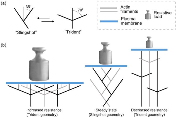
- Mueller J, Szep G, Nemethova M, de Vries I, Lieber AD, Winkler C, Kruse K, Small JV, Schmeiser C, Keren K, et al. Load adaptation of lamellipodial actin networks. Cell. 2017;171:188–200e116. Orientation of actin filaments in lamellipodia of migrating fish keratocytes is revealed by electron tomography of negatively stained cells. When plasma membrane tension was increased by pipet aspiration or decreased by severing off an attached cell region, actin filaments in lamellipodia reoriented from a conventional steady state orientation of ±35° relative to the direction of migration to angles that were close to 0° and ±70°. - PubMed
-
- Bieling P, Li TD, Weichsel J, McGorty R, Jreij P, Huang B, Fletcher DA, Mullins RD. Force feedback controls motor activity and mechanical properties of self-assembling branched actin networks. Cell. 2016;164:115–127. Single-molecule fluorescence microscopy was used to investigate in vitro assembly of branched actin networks growing as pillars from small micropatterned islands. Application of resistive loads using an AFM cantilever resulted in decreased growth velocity and increased network density without changes in the estimated average filament lengths. - PMC - PubMed
Publication types
MeSH terms
Substances
Grants and funding
LinkOut - more resources
Full Text Sources
Other Literature Sources
