

Comment
doi: 10.1016/j.jid.2017.11.002.
Shining a Light on Black Holes in Keratinocytes
Affiliations
- PMID: 29477191
- PMCID: PMC5832358
- DOI: 10.1016/j.jid.2017.11.002
Item in Clipboard
Comment
Shining a Light on Black Holes in Keratinocytes
J Invest Dermatol.
2018 Mar.
Abstract
The mechanisms by which melanins are transferred from melanocytes and stored within keratinocytes to generate skin pigmentation are hotly debated. Correia et al. and Hurbain et al. provide evidence that melanin cores of melanosomes are secreted from melanocytes and taken up and stored within non-degradative membranous organelles in keratinocytes in the basal epidermis of human skin.
Copyright © 2017 The Authors. Published by Elsevier Inc. All rights reserved.
Figures
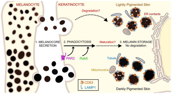
Melanocores are secreted by melanocytes into the extracellular space and phagocytosed by keratinocytes (step 1). PAR2 signaling and Rab5 are required for melanocore phagocytosis (step 2). Melanocores in keratinocytes are stored in non-degradative compartments bounded by single membranes that contain CD63 and LAMP2 and that contact rough and smooth ER, mitochondria, and small tubular membranes of unknown origin (step 3). These compartments harbor either clustered structures or dense, isolated structures in all skin types, but the ratio of clustered to isolated melanocore structures decreases as skin pigmentation increases (step 3, top vs. bottom). Although it is not yet clear how melanin storage organelles in keratinocytes mature, some clustered melanocores in lightly pigmented skin may undergo partial degradation (steps 2 to 3).
Comment on
-
Melanin Transferred to Keratinocytes Resides in Nondegradative Endocytic Compartments.J Invest Dermatol. 2018 Mar;138(3):637-646. doi: 10.1016/j.jid.2017.09.042. Epub 2017 Oct 24. J Invest Dermatol. 2018. PMID: 29074272
References
-
- Correia M, Moreiras H, Pereira F, Neto M, Festas T, Tarafder A, et al. Melanin transferred to keratinocytes resides in non-degradative compartments. J Invest Dermatol - PubMed
-
- Ebanks JP, Koshoffer A, Wickett RR, Schwemberger S, Babcock G, Hakozaki T, et al. Epidermal keratinocytes from light vs. dark skin exhibit differential degradation of melanosomes. J Invest Dermatol. 2011;131(6):1226–33. - PubMed
-
- Hurbain I, Romao M, Sextius P, Bourreau E, Marchal C, Bernerd F, et al. Melanosome distribution in keratinocytes in different skin types: melanosome clusters are not degradative organelles. J Invest Dermatol - PubMed
-
- Konrad K, Wolff K. Hyperpigmentation, melanosome size, and distribution patterns of melanosomes. Arch Dermatol. 1973;107(6):853–60. - PubMed
Publication types
MeSH terms
Substances
Grants and funding
LinkOut - more resources
Full Text Sources
Other Literature Sources
