. 2018 Apr 11:71:199-207.
doi: 10.1016/j.jbiomech.2018.02.007. Epub 2018 Feb 11.

Control of cellular adhesion and myofibroblastic character with sub-micrometer magnetoelastic vibrations

Affiliations

Control of cellular adhesion and myofibroblastic character with sub-micrometer magnetoelastic vibrations

Hal R Holmes et al. J Biomech. .

Abstract

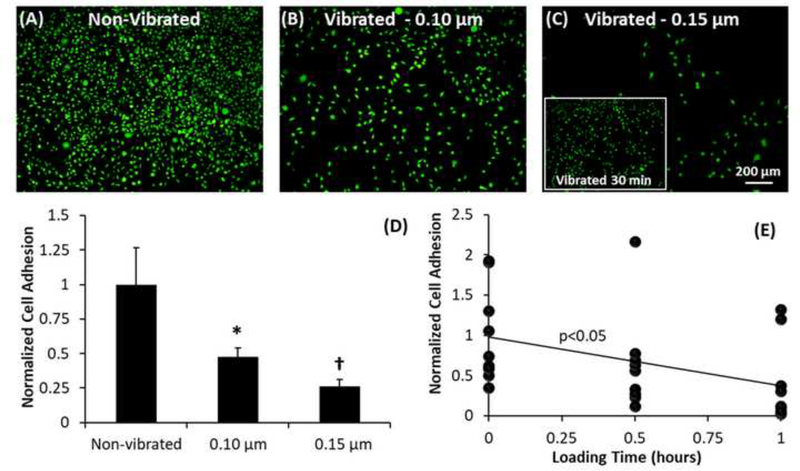
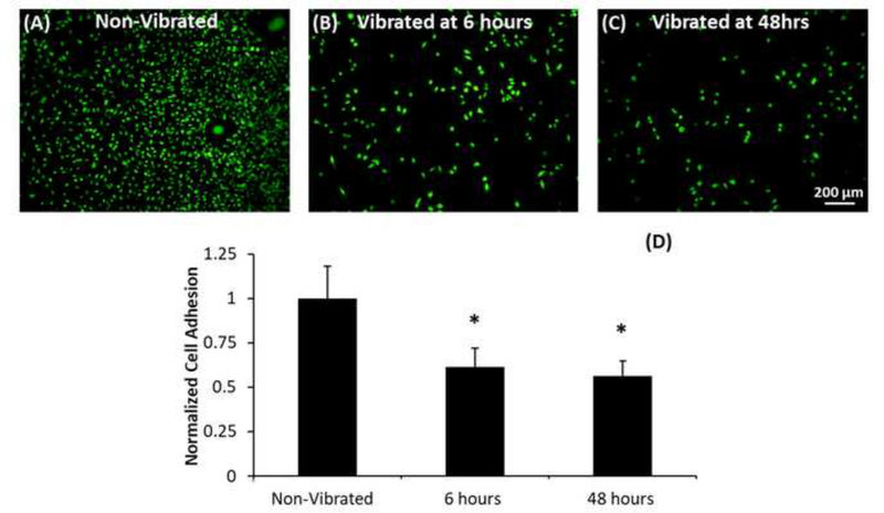
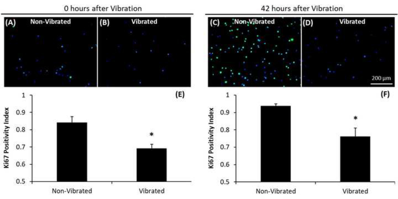
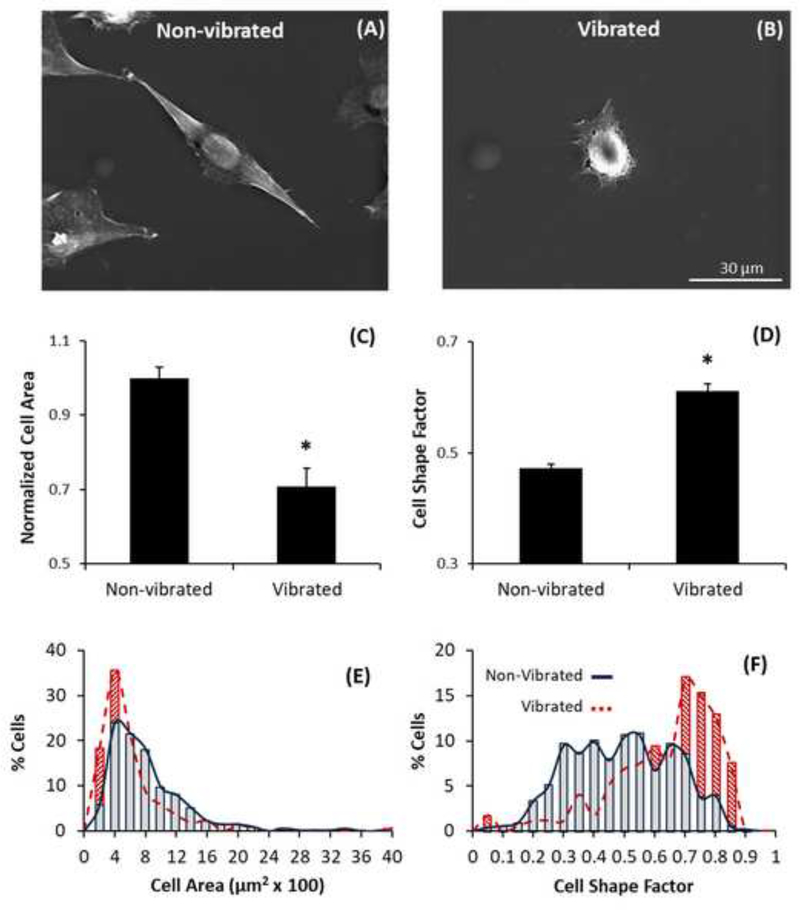
The effect of sub-cellular mechanical loads on the behavior of fibroblasts was investigated using magnetoelastic (ME) materials, a type of material that produces mechanical vibrations when exposed to an external magnetic AC field. The integration of this functionality into implant surfaces could mitigate excessive fibrotic responses to many biomedical devices. By changing the profiles of the AC magnetic field, the amplitude, duration, and period of the applied vibrations was altered to understand the effect of each parameter on cell behavior. Results indicate fibroblast adhesion depends on the magnitude and total number of applied vibrations, and reductions in proliferative activity, cell spreading, and the expression of myofibroblastic markers occur in response to the vibrations induced by the ME materials. These findings suggest that the subcellular amplitude mechanical loads produced by ME materials could potentially remotely modulate myofibroblastic activity and limit undesirable fibrotic development.

Keywords: Magnetoelastic; Mechanotransduction; Vibrations.


Conflict of interest statement

Conflict of Interest

The authors have no other conflicts of interest to declare.

Figures

Figure 1:
Figure 1:. Cell adhesion is dependent on vibration amplitude and loading duration.
Qualitative images of cell adhesion on ME materials that are non-vibrated (A), loaded with 0.10 μm (B), and 0.15 μm (C) vibrations. Inset shows cell adhesion after 30 minute loading with 0.15 μm vibrations. Quantitative assessment (D) shows significantly (p<0.05) different levels of cell adhesion on each loading group, demonstrating that the amplitude of applied vibrations can selectively control cell adhesion to a substrate. Regression analysis (E) shows significant (p<0.05) probability that cell adhesion is also dependent on duration of vibrational loading. *,† indicate statistically lower (p<0.05) levels of cell adhesion than non-vibrated controls and all other groups, respectively.
Figure 2:
Figure 2:. Cell adhesion is dependent on the vibration period (time between applied vibrations).
Qualitative images of cell adhesion on ME material surfaces that are non-vibrated (A), vibrated with a delay interval of 10 seconds (B), or vibrated with a delay interval of 1 second (C). Quantitative assessment of cell adhesion (D) shows significantly less cell adhesion on samples with a 1 second delay interval in comparison to non-vibrated controls, but no significant differences in cell adhesion were observed between any other groups. This result suggests that the period of sub-micron vibrations effects the response of adherent cells. Visual representation of differences in delay interval between loading groups (E). All vibrations were 0.15 μm in magnitude. * statistically significant (p<0.05) differences in cell adhesion compared to non-vibrated control.
Figure 3:
Figure 3:. Submicron ME vibrations can modulate cell adhesion during early or late stages of cell proliferation.
Fibroblasts cultured on non-vibrated ME materials (A) and ME materials loaded with submicron vibrations (0.15 μm; 160–170 kHz) 6 hours (B) or 48 hours (C) after seeding; all groups were stained and imaged after 48 hour vibration. Significant (p<0.05) differences in cell adhesion, quantified via direct cell counting, were observed between both loading groups and non-vibrated controls (D) but no significant differences in cell adhesion were observed between loading groups. This observation suggests that sub-micron vibrations may also effect proliferation behavior in addition to inducting immediately detachment. * indicate statistically significant differences (p<0.05) in cell adhesion compared to non-vibrated control.
Figure 4:
Figure 4:. ME vibrations reduce proliferative character of fibroblasts.
Cells, fixed with 4% paraformaldehyde immediately (A-B) or 42 hours (C-D) after loading application (0.15 μm from 150–170 kHz applied 6 hours after seeding), were stained using primary Ki-67 anti-body (Abcam) with Alexfluor-488 conjugated secondary anti-body (Abcam) and DAPI (Millipore) to visualize Ki-67 presence and cell nuclei, respectively. The Ki-67 positivity index (E-F), defined as the ratio of Ki-67 positive cells to total cells as determined by direct cell counting, was significantly different between non-vibrated and vibrated groups for both time points. * indicate statistically significant differences (p<0.05) in Ki-67positivity compared to non-vibrated controls.
Figure 5:
Figure 5:. ME vibrational loading induces a reduction in cell spreading.
Sub-micron vibrations (0.15 μm, 160–170 kHz) were applied 48 hours after seeding. Cells imaged on a JEOL JSM-6400 SEM (A-B) and measured for average area (C) and shape factor (D) using ImageJ software. Quantitative analysis showed significant differences in cell area and shape factor between vibrated and non-vibrated groups. The distribution of average cell area (E) and cell shape factor (F) is presented as a 20-bin histogram. * indicate statistically significant (p<0.05) differences in cell area and shape factor compared to non-vibrated control.
Figure 6:
Figure 6:. Changes in cytoskeletal organization, focal adhesion formation, and expression of TGF-β1 receptors occur in response to ME vibrations.
Following vibrational loading (applied 48 hours after seeding), cells were fixed and stained using phalloidin (Invitrogen) (Ai-Aii) and anti-vinculin antibody (Abcam) with FITC conjugated secondary (Abcam) (Aiii-Aiv) to mark the actin cytoskeleton and sites of focal adhesions (vinculin), respectively. Inset shows magnified view of actin fibers. Fluorescent images, quantified using ImageJ software, showed a significant reduction in average cell area (Av) and increase in cell shape factor (Avi). Western blot analyses was performed for vinculin (Avii), focal adhesion kinase (FAK; Avii), and TGF-β1 RII (Bi) using anti-vinculin (Abcam), anti-FAK (Abcam), and anti-TGF-β1 RII primary antibodies with HRP-conjugated secondary anti-bodies (Santa Cruz). Endogenous TGFβ−1 (Bii) as determined by ELISA assay demonstrates that sub-micron vibrations do not significantly affect the expression of TGFβ−1. Lysates were collected 24 hours (non-vibrated; Biii), 48 hours (non-vibrated and vibrated; Biv-v) and 72 hours (vibrated; Bvi) after seeding. Results show vinculin and FAK expression increase from 24 to 48 hours, decrease after vibrational loading, and increase again after loading (72 hours). TGF-β1 RII expression also increases from 24 to 48 hours and decreases after vibration, but remains low after loading. * indicate statistically significant differences (p<0.05) in cell area and shape factor compared to non-vibrated control.

Similar articles

Cited by

References

    1. Anderson JM, Rodriguez A, Chang DT, 2008. Foreign body reaction to biomaterials, in: Seminars in Immunology. Elsevier, pp. 86–100. - PMC - PubMed
    1. Cai QY, Cammers-Goodwin A, Grimes CA, 2000. A wireless, remote query magnetoelastic CO2 sensorElectronic Supplementary information available. See http://www.rsc.org/suppdata/EM/b0/b004929h. J. Environ. Monit 2, 556–560. - PubMed
    1. Cai QY, Grimes CA, 2000. A remote query magnetoelastic pH sensor. Sens. Actuators B Chem 71, 112–117. - PubMed
    1. Cai QY, Jain MK, Grimes CA, 2001. A wireless, remote query ammonia sensor. Sens. Actuators B Chem 77, 614–619. - PubMed
    1. Chen K-D, Li Y-S, Kim M, Li S, Yuan S, Chien S, Shyy JY, 1999. Mechanotransduction in response to shear stress roles of receptor tyrosine kinases, integrins, and Shc. J. Biol. Chem 274, 18393–18400. - PubMed

Publication types

LinkOut - more resources