

Identification of the Differential Expression Profiles of Serum and Tissue Proteins During Rat Hepatocarcinogenesis
- PMID: 29478368
- PMCID: PMC5833169
- DOI: 10.1177/1533034618756785
Identification of the Differential Expression Profiles of Serum and Tissue Proteins During Rat Hepatocarcinogenesis
Abstract
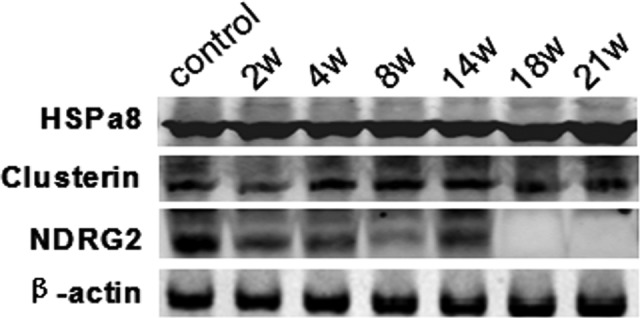
The pathogenesis of hepatocellular carcinoma is complex and not fully known yet. This study aims to screen and identify the differentially expressed proteins in peripheral blood and liver tissue samples from rat hepatocellular carcinoma and to further clarify the pathogenesis and discover the specific tumor markers and molecular targets of hepatocellular carcinoma. The hepatocellular carcinoma model of Wistar rats were induced by chemical carcinogen. The serum and liver tissue samples were obtained after induction for 2, 4, 8, 14, 18, and 21 weeks. The results showed that the clusterin (IPI00198667), heat shock protein a8 (IPI00208205), and N-myc downstream-regulated gene-2 (IPI00382069) being closely related to hepatocarcinogenesis were eventually identified from the 30 different proteins. As the time progressed, the serum levels of clusterin and heat shock protein a8 increased gradually during induced liver cancer in rats. However, the serum N-myc downstream-regulated gene 2 level in induced liver cancer in rats underwent biphasic changes, and the serum N-myc downstream-regulated gene 2 level decreased at the 8th week, increased at the 14th week, and then decreased significantly. Statistical difference occurred in protein expression of clusterin and heat shock protein a8 in liver tissues at the different time points. In the liver tissues, the N-myc downstream-regulated gene 2 level decreased gradually at the 8th week, increased gradually at the 14th week, and then decreased significantly after 14 weeks. The study demonstrated that heat shock protein a8, clusterin, and N-myc downstream-regulated gene 2 participated in the process of abnormal cell division, proliferation, and carcinogenesis of liver cells during hepatocarcinogenesis.
Keywords: diagnosis; hepatocellular carcinoma (HCC); occurrence; proteomics; target therapy.
Conflict of interest statement
Figures


Similar articles
-
Combined serum and tissue proteomic study applied to a c-Myc transgenic mouse model of hepatocellular carcinoma identified novel disease regulated proteins suitable for diagnosis and therapeutic intervention strategies.J Proteome Res. 2011 Jul 1;10(7):3012-30. doi: 10.1021/pr101207t. Epub 2011 Jun 6. J Proteome Res. 2011. PMID: 21644509
-
Gene expression profiling of MYC-driven tumor signatures in porcine liver stem cells by transcriptome sequencing.World J Gastroenterol. 2015 Feb 21;21(7):2011-29. doi: 10.3748/wjg.v21.i7.2011. World J Gastroenterol. 2015. PMID: 25717234 Free PMC article.
-
An Immune Gene Expression Signature Associated With Development of Human Hepatocellular Carcinoma Identifies Mice That Respond to Chemopreventive Agents.Gastroenterology. 2019 Nov;157(5):1383-1397.e11. doi: 10.1053/j.gastro.2019.07.028. Epub 2019 Jul 22. Gastroenterology. 2019. PMID: 31344396 Free PMC article.
-
Role of epigenetic aberrations in the development and progression of human hepatocellular carcinoma.Cancer Lett. 2014 Jan 28;342(2):223-30. doi: 10.1016/j.canlet.2012.01.038. Epub 2012 Feb 2. Cancer Lett. 2014. PMID: 22306342 Free PMC article. Review.
-
Role of DLC1 tumor suppressor gene and MYC oncogene in pathogenesis of human hepatocellular carcinoma: potential prospects for combined targeted therapeutics (review).Int J Oncol. 2012 Aug;41(2):393-406. doi: 10.3892/ijo.2012.1474. Epub 2012 May 10. Int J Oncol. 2012. PMID: 22580498 Free PMC article. Review.
Cited by
-
Evaluating the diagnostic accuracy of heat shock proteins and their combination with Alpha-Fetoprotein in the detection of hepatocellular carcinoma: a meta-analysis.BMC Gastroenterol. 2024 May 21;24(1):178. doi: 10.1186/s12876-024-03260-5. BMC Gastroenterol. 2024. PMID: 38773451 Free PMC article.
-
The Influence of Clusterin Glycosylation Variability on Selected Pathophysiological Processes in the Human Body.Oxid Med Cell Longev. 2022 Aug 28;2022:7657876. doi: 10.1155/2022/7657876. eCollection 2022. Oxid Med Cell Longev. 2022. PMID: 36071866 Free PMC article. Review.
-
Role of secretory clusterin in hepatocarcinogenesis.Transl Gastroenterol Hepatol. 2018 Aug 1;3:48. doi: 10.21037/tgh.2018.07.13. eCollection 2018. Transl Gastroenterol Hepatol. 2018. PMID: 30221206 Free PMC article. Review.
References
-
- Maluccio M, Covey A. Recent progress in understanding, diagnosing, and treating hepatocellular carcinoma. CA Cancer J Clin. 2012;62(6):394–399. - PubMed
-
- Jian ZX. Progress and prospect in the treatment of primary liver cancer. J Pract Med. 2007;23(6):773–775.
-
- Ren F. Research progress of proteomics on nonmalignant liver diseases with the application of MALDI-TOF-M. J Pract Hepatol. 2009;12(1):59–63.
-
- Shen YC, Yao DF. Evaluation of proteomics technology in early diagnosis and monitoring of Hepatocellular Carcinoma. J Med Res. 2008;37(9):12–15.
Publication types
MeSH terms
Substances
LinkOut - more resources
Full Text Sources
Other Literature Sources
Medical
Research Materials
