Review
. 2018 Feb;39(2):200-208.
doi: 10.1016/j.tips.2017.11.009. Epub 2018 Jan 28.

Subcellular Organization of GPCR Signaling

Affiliations
Review

Subcellular Organization of GPCR Signaling

Kelsie Eichel et al. Trends Pharmacol Sci. 2018 Feb.

Abstract

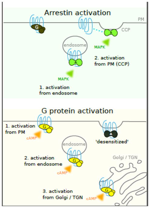
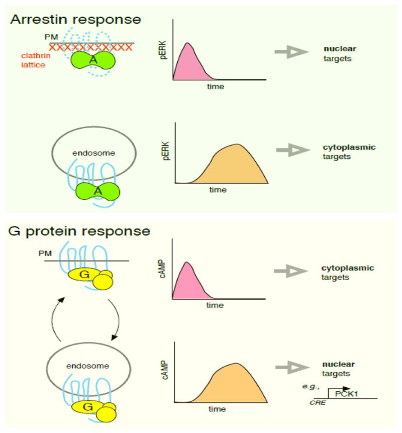
G protein-coupled receptors (GPCRs) comprise a large and diverse class of signal-transducing receptors that undergo dynamic and isoform-specific membrane trafficking. GPCRs thus have an inherent potential to initiate or regulate signaling reactions from multiple membrane locations. This review discusses emerging insights into the subcellular organization of GPCR function in mammalian cells, focusing on signaling transduced by heterotrimeric G proteins and β-arrestins. We summarize recent evidence indicating that GPCR-mediated activation of G proteins occurs not only from the plasma membrane (PM) but also from endosomes and Golgi membranes and that β-arrestin-dependent signaling can be transduced from the PM by β-arrestin trafficking to clathrin-coated pits (CCPs) after dissociation from a ligand-activated GPCR.

Keywords: G protein; GPCR; Golgi; arrestin; endosome; signaling.


Conflict of interest statement

Conflict of Interest

The authors declare no conflict of interest.

Figures

Figure 1
Figure 1
Schematic summarizing subcellular locations of GPCR-mediated activation of arrestins and heterotrimeric G proteins supported by data discussed in the text. Top panel: Arrestin (β-arrestins -1 and -2) are thought to be subject to activation both in endosomes and the PM. Bottom panel: Gs activation by various GPCRs (see text for details) has been described in the PM, endosomes, Golgi apparatus and trans-Golgi network.
Figure 2
Figure 2
Schematic contrasting current view of functional impact of localized signaling transduced by β-arrestins and Gs. Top panel: GPCR-mediated activation of β-arrestins in the PM is thought to produce a rapid ERK response that can access nuclear targets. Activation in endosomes produces delayed ERK activation and sequesters ERK in the cytoplasm, thereby preventing access to nuclear targets. Bottom panel: GPCR-mediated activation of Gs in the PM produces a rapid and transient response that activates cAMP-dependent targets in the PM and cytoplasm. Activation in endosomes produces a delayed response that preferentially accesses nuclear targets (induction of the cAMP-responsive gene PCK1 is shown as an example. For GPCRs that are capable of efficient recycling, PM and endosome-initiated activation waves can occur repeatedly in the prolonged presence of agonist through multiple rounds of endocytosis and recycling (depicted as curved arrows).

Similar articles

Cited by

References

    1. Di Fiore PP, von Zastrow M. Endocytosis, signaling, and beyond. Cold Spring Harb Perspect Biol. 2014:6. - PMC - PubMed
    1. Irannejad R, von Zastrow M. GPCR signaling along the endocytic pathway. Curr Opin Cell Biol. 2014;27:109–116. - PMC - PubMed
    1. Irannejad R, et al. Effects of endocytosis on receptor-mediated signaling. Curr Opin Cell Biol. 2015;35:137–143. - PMC - PubMed
    1. Ni Q, et al. Live-cell imaging of cell signaling using genetically encoded fluorescent reporters. FEBS J. 2017 doi: 10.1111/febs.14134. - DOI - PMC - PubMed
    1. Rasmussen SGF, et al. Structure of a nanobody-stabilized active state of the beta2 adrenoceptor. 2011 - PMC - PubMed

Substances