. 2018 Sep 15;84(6):401-412.
doi: 10.1016/j.biopsych.2018.01.019. Epub 2018 Feb 2.

Dopamine D1 Receptor-Positive Neurons in the Lateral Nucleus of the Cerebellum Contribute to Cognitive Behavior

Affiliations

Dopamine D1 Receptor-Positive Neurons in the Lateral Nucleus of the Cerebellum Contribute to Cognitive Behavior

Timothy M Locke et al. Biol Psychiatry. .

Abstract

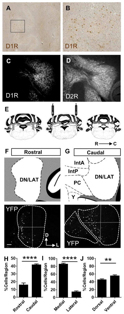
Background: Studies in humans and nonhuman primates have identified a region of the dentate nucleus of the cerebellum, or the lateral cerebellar nucleus (LCN) in rodents, activated during performance of cognitive tasks involving complex spatial and sequential planning. Whether such a subdivision exists in rodents is not known. Dopamine and its receptors, which are implicated in cognitive function, are present in the cerebellar nuclei, but their function is unknown.

Methods: Using viral and genetic strategies in mice, we examined cellular phenotypes of dopamine D1 receptor-positive (D1R+) cells in the LCN with whole-cell patch clamp recordings, messenger RNA profiling, and immunohistochemistry to examine D1R expression in mouse LCN and human dentate nucleus of the cerebellum. We used chemogenetics to inhibit D1R+ neurons and examined behaviors including spatial navigation, social recognition memory, prepulse inhibition of the acoustic startle reflex, response inhibition, and working memory to test the necessity of these neurons in these behaviors.

Results: We identified a population of D1R+ neurons that are localized to an anatomically distinct region of the LCN. We also observed D1R+ neurons in human dentate nucleus of the cerebellum, which suggests an evolutionarily conserved population of dopamine-receptive neurons in this region. The genetic, electrophysiological, and anatomical profile of mouse D1R neurons is consistent with a heterogeneous population of gamma-aminobutyric acidergic, and to a lesser extent glutamatergic, cell types. Selective inhibition of D1R+ LCN neurons impairs spatial navigation memory, response inhibition, working memory, and prepulse inhibition of the acoustic startle reflex.

Conclusions: Collectively, these data demonstrate a functional link between genetically distinct neurons in the LCN and cognitive behaviors.

Keywords: Cerebellar nuclei; Cerebellum; Cognition; DREADD receptor; Dopamine D(1) receptor; RiboTag.


Figures

Figure 1
Figure 1
Immunohistochemistry reveals staining for D1R in human DCN and mouse LCN. Mapping reveals subregional localization of D1R neurons in LCN. A, Immunohistochemistry for the D1R protein in human DCN (in a parasagittal section), square in I is zone at higher magnification in B. Staining for D1R was positive in each of 5 cases. C, Staining of D1R in the green color channel in lateral nucleus of the cerebellum in a coronal section. D, Staining of D2R in the red color channel in lateral nucleus of the cerebellum in a coronal section. E, Illustration depicting the lateral (dentate) nucleus (DN/LAT) from the rostral to caudal extent that was analyzed for D1R neuron location. F, Illustration of the rostral region of LCN in coronal plane (top), representative image of D1R:Tmto expression in rostral LCN overlaid with divisions of dorsal, ventral, medial and lateral zones (bottom). Dorsal and lateral orientations are denoted in inset. Scale = 120 μm. G, Illustration of the caudal region of LCN in coronal plane (top), representative image of D1R:Tmto expression in caudal LCN overlaid with divisions of dorsal, ventral, medial and lateral zones (bottom), In addition to the LCN, few cells were observed in the parvocellular region of the LCN and the interposed nuclei. (DN = Dentate (or Lateral) Nucleus; IntA = Interposed Nucleus Anterior part; IntP: Interposed Nucleus, posterior part; PC: Dentate (or Lateral) Nucleus, parvocellular part; Y: Nucleus Y of the vestibular complex). H, Quantification of distribution of D1R:YFP positive cells in rostral vs. caudal zones (t=7.42, df=29). I, Quantification of distribution of D1R:GFP positive cells in medial vs. lateral zones (t=19.2, df=56). J, Quantification of distribution of D1R:GFP positive cells in dorsal vs. ventral zones (t=2.83, df=54). Results were acquired from 4 mice, 4 sections per mouse counted bilaterally and are represented as the average number of cells per section per side or the percentage of cells within each region per section per side. Illustrations in A–C are from Paxinos and Franklin, 2013. ** P < 0.01, **** P < 0.0001, Student’s t test, two-tailed.
Figure 2
Figure 2
Electrophysiological characterization of D1R LCN neurons in mice. A, Average AP waveforms of Type I and Type II neurons, Scale = 20 mV, 2 ms. B, AP Threshold, AP Peak, and AHP Peak in Type I and Type II neurons. C, Time to AHP peak in Type I and Type II neurons. ***P < 0.001, Student’s t test, two-tailed, t=4.04, df=24. D, AP half-width of Type I and Type II neurons. **** P < 0.0001, Student’s t test, two-tailed, t=5.33, df=24. E, Example Type I (top) and Type II (middle) neurons before, during, and after 50 pA current injection (bottom), Scale = 20 mV, 200 ms. F, Capacitance of Type I and Type II neurons. G–L: Type I N = 16; Type II N = 10, **** P < 0.0001, Student’s t test, two-tailed, t=5.87, df=24. G, Distribution of measured surface areas of D1R neurons in the DNC revealing a non-normal distribution (N = 306 cells, Shapiro-Wilk, P < 0.05).
Figure 3
Figure 3
Translational profiling and immunohistochemistry reveal identities of D1R LCN neurons in mice. A, Expression of Rpl22-HA in D1R neurons, Scale bar = 60 μm (inset scale bar = 10 μm), in the LCN. B, Schematic of RiboTag methodology: Following cell lysis (1), HA antibody-coupled magnetic beads immuno-isolate tagged polysomes and associated mRNA (2). mRNA are isolated (3) and cDNA is generated from both input and immunoprecipitated mRNA. C, qRT-PCR analysis of immunoprecipitate relative to input demonstrating significant enrichment of Drd1a, Drd2, Vgat, Gad1, Glyt2, and Penk (enriched markers in black), relative to Cnp, an oligodendrocyte marker in D1R cells of LCN. Drd3, encoding the dopamine D3 receptor, and oligodendroglial marker (Cnp) were de-enriched (white), while the marker of glutamatergic neurons (Vglut2), was neither enriched or de-enriched (grey). ****P < 0.0001, ** P < 0.01, *P < 0.05, One-way ANOVA, n = 3 pooled samples of 7 mice/pool (F9, 20 = 15.3). D, qRT-PCR analysis of immunoprecipitate relative to input demonstrating significant enrichment of Drd2, Penk, Glyt2, Gad1, and Vgat (enriched markers in black), relative to Cnp, in Vgat+ cells of LCN. Cnp, Vglut2, and Drd3 were de-enriched (white), while Drd1a was neither enriched or de-enriched (grey). ****P < 0.0001, ** P < 0.01, *P < 0.05, One-way ANOVA, n = 4 pooled samples of 4 mice/pool (F8,32 = 20.1). E, qRT-PCR analysis of immunoprecipitate relative to input demonstrating significant enrichment of Drd3, in Vglut2+ cells (black). Glyt2, Gad1, and Vgat, were de-enriched (white), while Drd1a, Drd2, and Penk, were neither enriched nor de-enriched (grey). ****P < 0.0001, ** P < 0.01, *P < 0.05, One-way ANOVA, n = 4 samples of 1 mouse/sample (F8,32 = 23.3). F, Venn diagram illustrating distribution of D1R expression in neural subtypes residing in the LCN.
Figure 4
Figure 4
A–P, Projection patterns of D1R LCN neurons in D1R-Cre mice compared with Vgat-Cre and Vglut2-Cre mice. A, Illustration depicting the lateral dentate nucleus (DN/LAT) location of injection for Synapto-GFP and mCherry viral constructs (B, C) depicted in D–G. B, Illustration of Synapto-GFP viral construct. C, Illustration of mCherry viral construct. D–F, Histochemistry demonstrating synaptophysin-GFP (Syn-GFP) expression in DNC of Drd1aCre/+ VgatCre/+, and Vglut2Cre/+ mice, respectively; scale bar represents 60 μm. G–I, mCherry expressing neurons are observed in the cerebellar cortex along with a number of Syn-GFP puncta. Scale bar represents 200 μm. J–O, Nucleocortical rosette-like or Bead-like synapses in cerebellar cortex in each of three strains of mice. Scale bar represents 2 μm.
Figure 5
Figure 5
DREADD Receptor expression in D1R LCN neurons and CNO application results in altered performance on the Barnes Maze. A, Schema for when CNO was injected IP prior to training and probe tests above performance of each group during training trials, as measured by average duration in the goal quadrant (left) and as measured by nose pokes in target quadrant holes and goal hole (right). N = 17 D1R:Hm4Di mice, and N = 23 littermate D1R:GFP control mice. For goal quadrant duration, Error bars are SEM. * P < 0.05 (Two-way rmANOVA, 2 factors were significant: Interaction between training and presence of Hm4Di (F3,114 = 3.14, *P < 0.05) and training (F3,114 = 5.3, **P < 0.01). the factor of presence of Hm4Di alone was not significant. Holm-Sidak’s post hoc multiple comparisons test did not indicate any difference in target quadrant duration for any days of training. For nose pokes in goal quadrant, (Two-way rmANOVA) 2 factors were significant: Interaction between training and presence of Hm4Di (F3,114 = 3.92, *P < 0.05) and Training (F3,114 = 8.49, *P < 0.0001); The factor of presence of Hm4Di alone was not significant. Holm-Sidak’s post hoc multiple comparisons test indicated that the difference in nose pokes between groups during training was only significantly different on day 4 of training, *P < 0.05, DF =152. B, Performance of each group during memory recall during the probe trial of the Barnes maze, as measured by nose pokes in target quadrant holes. N = 17, D1R:Hm4Di mice and N = 23 littermate D1R:GFP control mice. Error bars are SEM. * P < 0.05 Unpaired Student’s t test, two tailed, t=2.52, df=38. C, Performance of each group during memory recall during the probe trial of the Barnes maze, as measured by nose pokes in goal hole. D1R:GFP had significantly more nose pokes in goal hole than D1R:Hm4Di mice on probe trial, * P < 0.05, Unpaired Student’s t test, two tailed, t=2.23, df=38. D, Representative path traces of D1R:GFP after IP injection of CNO and E, D1R:Hm4Di after IP injection of CNO on the probe trial on Day 5 of the Barnes Maze. F, Social approach as measured by time spent in arena with a novel mouse or novel object. Both groups preferred social interaction (F1,72 = 48.52, P < 0.0001). (N = 22, D1R:GFP mice, and N= 17, D1R:Hm4Di mice, Two-way ANOVA). No significant differences were found for presence of Hm4Di or interaction (presence of Hm4Di X zone). G, Social preference as measured by time spent in arena with a novel mouse or familiar mouse. Factors for interaction (Presence of Hm4Di X zone, F1,72 = 4.59, *P < 0.05) and zone (F1,72 = 4.19, P < 0.05) were significant (N = 22, D1R:GFP mice, and N= 17, D1R:Hm4Di mice, **P < 0.01, Two-way ANOVA, Sidak’s multiple comparisons test). H, Prepulse inhibition of the acoustic startle reflex (N = 22, D1R:GFP mice, and N= 17, D1R:Hm4Di mice), was significantly different for presence of Hm4Di (F1,37 = 6.95, *P < 0.05) and dB of prepulse, P < 0.0001, F2,74 = 44.22), but not interaction, Two-way rmANOVA.
Figure 6
Figure 6
Peak Interval Timing and Working Memory are regulated by D1R neurons in the LCN. A and B, Schematics describing contingencies of DRL operant timing task. C, Distribution of latencies of lever presses after reward in the DRL-10 for D1R:GFP control mice (black circles) and D1R:Hm4Di mice (red diamonds). Group means ± SEM are presented for latencies binned in 2s intervals. Dotted line represents minimum response latency that is rewarded for the DRL paradigm (2-way RM ANOVA, Presence of Hm4Di x time interaction: Week 3: F29,261 = 5.61, P < 0.0001; N = 6, D1R:Hm4Di mice and N = 5, D1R:GFP mice). Inset is the mean peak of response distribution of each group for this week of training in this 10 second DRL paradigm. D, Schematic describing the delayed alternation operant task. E–J, Performance of D1R:GFP control mice (black lines) and D1R:Hm4Di mice (red lines) on increasingly difficult versions of the task as measured by proportion correct (E–G) and pellets rewarded (H–J). Group means ± SEM are presented in E–J. E, 2 second delay, only significant factor was for training: F5,140 = 101.5, P < 0.0001; no significant differences between groups, or interaction between training and presence of Hm4Di. N = 12, D1R:Hm4Di mice and N = 18, D1R:GFP mice. F, 8 second delay, significant differences for factors of training: F4,112 = 16.16, P < 0.0001, and between groups, F1,28 = 7.2, *P < 0.05, but not interaction. N = 12, D1R:Hm4Di mice and N = 18, D1R:GFP mice. G, 16-second delay, factor of presence of Hm4Di was significant: F1,28 = 4.83, *P < 0.05. Interaction between groups and training were not significant. N = 12, D1R:Hm4Di mice and N = 18, D1R:GFP mice. H, 2 second delay, only significant factor was for training: F5,140 = 50.92, P < 0.0001; no significant differences between groups or interaction between presence of training and presence of Hm4Di. N = 12, D1R:Hm4Di mice and N = 18, D1R:GFP mice. I, 8 second delay, no significant differences for factors of training, Hm4Di groups, or interaction between groups and training were found. N = 12, D1R:Hm4Di mice and N = 18, D1R:GFP mice. J, 16-second delay, factor of presence of Hm4Di was significant: F1,28 = 4.35, *P < 0.05. Interaction between groups and training was not significant. Two-way rmANOVA N = 12, D1R:Hm4Di mice and N = 18, D1R:GFP mice.

Similar articles

Cited by

References

    1. Kawato M, Gomi H. A computational model of four regions of the cerebellum based on feedback-error learning. Biological cybernetics. 1992;68:95–103. - PubMed
    1. Medina JF, Nores WL, Ohyama T, Mauk MD. Mechanisms of cerebellar learning suggested by eyelid conditioning. Current opinion in neurobiology. 2000;10:717–724. - PubMed
    1. Raymond JL, Lisberger SG. Neural learning rules for the vestibulo-ocular reflex. J Neurosci. 1998;18:9112–9129. - PMC - PubMed
    1. Braak E, Arai K, Braak H. Cerebellar involvement in Pick’s disease: affliction of mossy fibers, monodendritic brush cells, and dentate projection neurons. Experimental neurology. 1999;159:153–163. - PubMed
    1. Parisi MA. Clinical and molecular features of Joubert syndrome and related disorders. American journal of medical genetics Part C, Seminars in medical genetics. 2009;151C:326–340. - PMC - PubMed

Publication types

Substances