. 2018 Mar 14;23(3):297-301.e4.
doi: 10.1016/j.chom.2018.01.006. Epub 2018 Feb 22.

Dampened STING-Dependent Interferon Activation in Bats

Affiliations

Dampened STING-Dependent Interferon Activation in Bats

Jiazheng Xie et al. Cell Host Microbe. .

Abstract

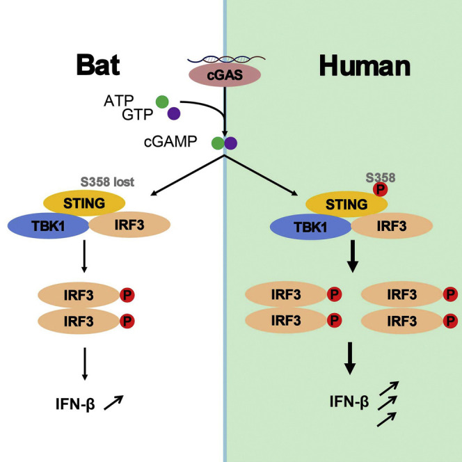
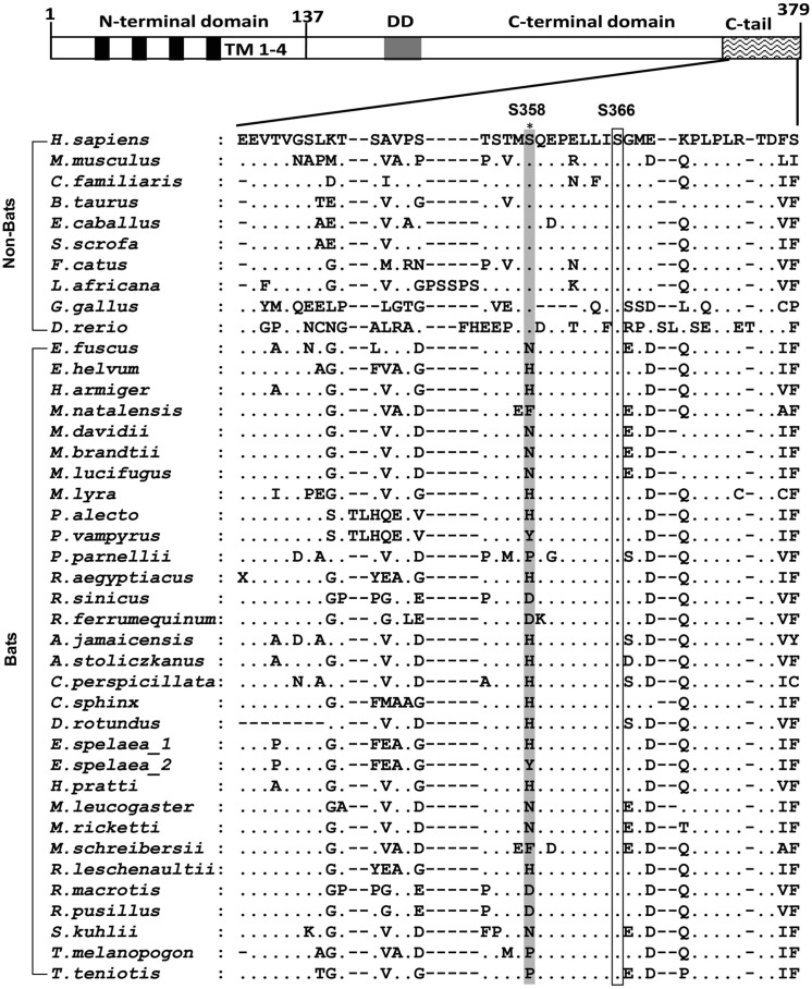
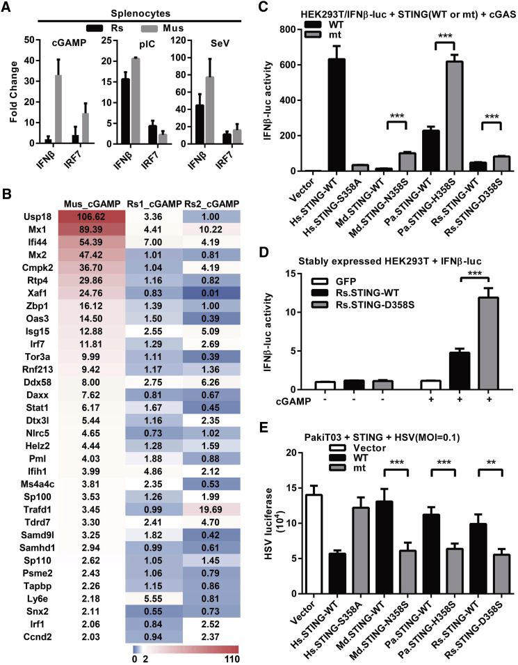
Compared with terrestrial mammals, bats have a longer lifespan and greater capacity to co-exist with a variety of viruses. In addition to cytosolic DNA generated by these viral infections, the metabolic demands of flight cause DNA damage and the release of self-DNA into the cytoplasm. However, whether bats have an altered DNA sensing/defense system to balance high cytosolic DNA levels remains an open question. We demonstrate that bats have a dampened interferon response due to the replacement of the highly conserved serine residue (S358) in STING, an essential adaptor protein in multiple DNA sensing pathways. Reversing this mutation by introducing S358 restored STING functionality, resulting in interferon activation and virus inhibition. Combined with previous reports on bat-specific changes of other DNA sensors such as TLR9, IFI16, and AIM2, our findings shed light on bat adaptation to flight, their long lifespan, and their unique capacity to serve as a virus reservoir.

Keywords: DNA sensing; STING; bats; dampened; interferon; virus.


Figures

None
Graphical abstract
Figure 1
Figure 1
Mutations of S358 in Bat STING STING domains are illustrated on top of the alignment. The highly conserved regions are boxed. Residue 358 is highlighted in gray. There are two Eonycteris spelaea STING sequences because of the polymorphism at residue 358. The full species name and accession numbers of STING or SRA data are listed in Table S1. See also Figure S1 and Table S1.
Figure 2
Figure 2
Dampened IFN Activation and Virus Inhibition by Bat STING (A) Splenocytes of Rhinolophus sinicus bats and mice (n = 3 cells from 3 animals each) were transfected with cGAMP (1 μg/mL) or poly I:C (1 μg/mL), or infected with SeV (100 hemagglutinin units/mL). Six hours later, the induction of IFNβ and IRF7 genes was determined by qPCR. Primers can be found in Table S2. (B) Transcriptome next-generation sequencing of splenocyte RNAs. The differentially expressed genes (DEGs) were analyzed by RSEM at FDR (false discovery rate) < 0.05. The ISG in the DEG sets of mice and Rs bat are listed. Fold change is indicated in color from 0 to 110. (C) Restoration of STING function by introducing S358 in bat STING. HEK293T cells were co-transfected with STING, cGAS, IFNβ promoter firefly luciferase, and renilla luciferase plasmids. Luciferase activity was determined 24 hr post-transfection. The blots showing protein levels can be found in Figure S2. (D) cGAMP treatment of HEK293T stably expressing various STING. Cells stably expressing the indicated proteins were transfected with IFNβ promoter firefly luciferase and renilla luciferase plasmids. Six hours later, cells were permeabilized in digitonin buffer with or without 1 μg/mL cGAMP. Luciferase activity was determined 16 hr after treatments. (E) PaKiT03 cells were transfected with indicated STING plasmids followed by infection with HSV-luciferase at MOI = 0.1 at 24 hr post-transfection. At 24 hr post-infection, HSV replication was determined by luciferase activity. Data from (A), (C), (D), and (E) are presented as the means ± SD, n = 3, ∗∗p < 0.01, ∗∗∗p < 0.001 (Student's t test). For (C) and (D), data represent fold change according to wells transfected with empty vector (set as 1). WT, wild-type; mt, mutant; Hs, Homo sapiens; Md, Myotis davidii; Pa, Pteropus alecto; Rs, Rhinolophus sinicus; pIC, poly I:C. See also Figure S2 and Table S2.

Similar articles

Cited by

References

    1. Ahn M., Cui J., Irving A.T., Wang L.F. Unique loss of the PYHIN gene family in bats amongst mammals: implications for inflammasome sensing. Sci. Rep. 2016;6:21722. - PMC - PubMed
    1. Barber G.N. STING: infection, inflammation and cancer. Nat. Rev. Immunol. 2015;15:760–770. - PMC - PubMed
    1. Barzilai A., Rotman G., Shiloh Y. ATM deficiency and oxidative stress: a new dimension of defective response to DNA damage. DNA Repair. 2002;1:3–25. - PubMed
    1. Brook C.E., Dobson A.P. Bats as 'special' reservoirs for emerging zoonotic pathogens. Trends Microbiol. 2015;23:172–180. - PMC - PubMed
    1. Channappanavar R., Fehr A.R., Vijay R., Mack M., Zhao J., Meyerholz D.K., Perlman S. Dysregulated type I interferon and inflammatory monocyte-macrophage responses cause lethal pneumonia in SARS-CoV-infected mice. Cell Host Microbe. 2016;19:181–193. - PMC - PubMed

MeSH terms