Case Reports
. 2018 Mar 1;102(3):494-504.
doi: 10.1016/j.ajhg.2018.01.020. Epub 2018 Feb 22.

Biallelic Mutations in ATP5F1D, which Encodes a Subunit of ATP Synthase, Cause a Metabolic Disorder

Collaborators, Affiliations
Case Reports

Biallelic Mutations in ATP5F1D, which Encodes a Subunit of ATP Synthase, Cause a Metabolic Disorder

Monika Oláhová et al. Am J Hum Genet. .

Abstract

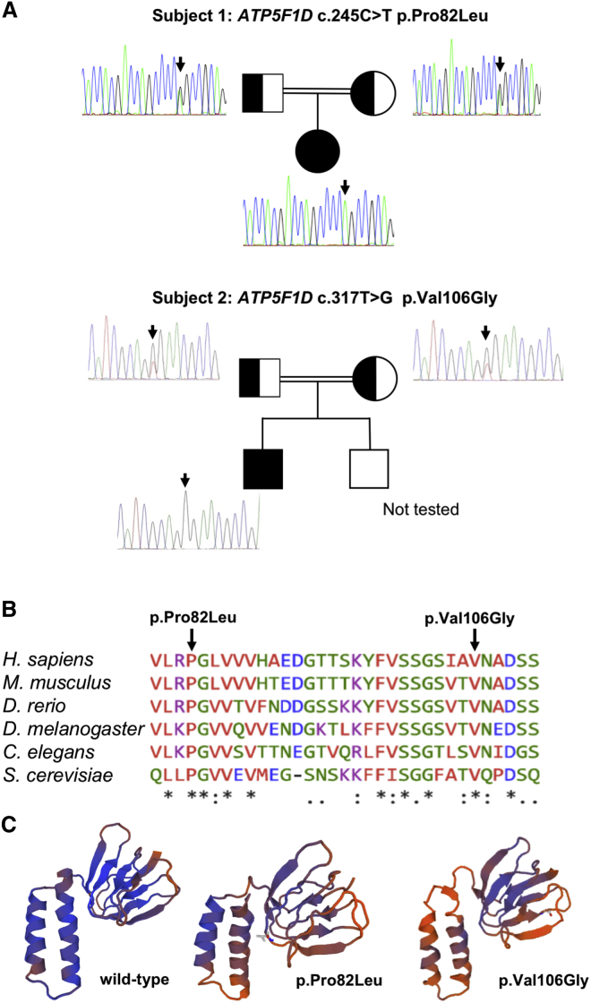
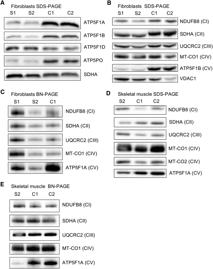
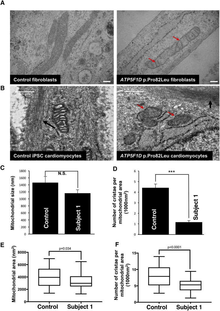
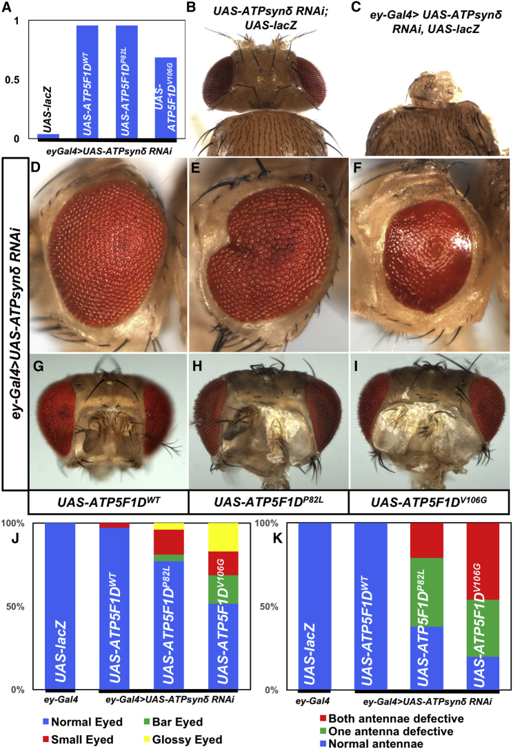
ATP synthase, H+ transporting, mitochondrial F1 complex, δ subunit (ATP5F1D; formerly ATP5D) is a subunit of mitochondrial ATP synthase and plays an important role in coupling proton translocation and ATP production. Here, we describe two individuals, each with homozygous missense variants in ATP5F1D, who presented with episodic lethargy, metabolic acidosis, 3-methylglutaconic aciduria, and hyperammonemia. Subject 1, homozygous for c.245C>T (p.Pro82Leu), presented with recurrent metabolic decompensation starting in the neonatal period, and subject 2, homozygous for c.317T>G (p.Val106Gly), presented with acute encephalopathy in childhood. Cultured skin fibroblasts from these individuals exhibited impaired assembly of F1FO ATP synthase and subsequent reduced complex V activity. Cells from subject 1 also exhibited a significant decrease in mitochondrial cristae. Knockdown of Drosophila ATPsynδ, the ATP5F1D homolog, in developing eyes and brains caused a near complete loss of the fly head, a phenotype that was fully rescued by wild-type human ATP5F1D. In contrast, expression of the ATP5F1D c.245C>T and c.317T>G variants rescued the head-size phenotype but recapitulated the eye and antennae defects seen in other genetic models of mitochondrial oxidative phosphorylation deficiency. Our data establish c.245C>T (p.Pro82Leu) and c.317T>G (p.Val106Gly) in ATP5F1D as pathogenic variants leading to a Mendelian mitochondrial disease featuring episodic metabolic decompensation.

Keywords: 3-methylglutaric aciduria; ATP synthase; complex V; exome sequencing; fibroblast; hyperammonemia; lactic acidosis; mitochondrial disease; model organism; oxidative phosphorylation.


Figures

Figure 1
Figure 1
Molecular Genetic Studies of ATP5F1D Variants (A) Pedigrees and sequencing chromatograms of the two affected families show segregation of the homozygous ATP5F1D variant c.245C>T (p.Pro82Leu) in subject 1 and c.317T>G (p.Val106Gly) in subject 2. (B) Multiple-sequence alignment confirms evolutionary conservation of p.Pro82Leu and p.Val106Gly in both human and flies. (C) SWISS-MODEL-predicted structure of wild-type, p.Pro82Leu, and p.Val106Gly ATP5F1D.
Figure 2
Figure 2
Biallelic Variants in ATP5F1D Impair the Steady-State Amounts of the F1FO ATP Synthase Complex and Subunits Immunoblot and BN-PAGE analysis were carried out on subject cultured skin fibroblasts and skeletal muscle samples as previously described., , SDS-PAGE and immunoblot analysis of whole-cell lysates (40 μg) isolated from cultured skin fibroblasts of affected subjects 1 (S1) and 2 (S2) and age-matched control individuals show (A) the steady-state amounts of complex V subunits (ATP5F1A, ATP5F1B, ATP5F1D, and ATP5PO) and (B) the amounts of individual OXPHOS complex subunits. One-dimensional BN-PAGE analysis was performed for assembled OXPHOS complexes in n-dodecyl-β-D-maltoside (DDM; 850520P, Sigma)-solubilized mitochondrial extracts isolated from control, S1, and S2 fibroblasts (C). Steady-state amounts (D) and assembly (E) of OXPHOS complexes and subunits in DDM-solubilized mitochondrial extracts from control and subject 2 skeletal muscle demonstrate a decrease in complex V. In (C) and (E), mitochondrial lysates (100 μg) were loaded on a 4%–16% native gel (Life Technologies), and then protein complexes were immobilized onto polyvinylidene difluoride membranes and subjected to immunoblotting with the indicated OXPHOS-subunit-specific antibodies. In (A)–(E), nuclear-encoded SDHA (ab14715, Abcam) or porin (VDAC1, ab14734, Abcam) was used as a loading control. Abbreviations are as follows: BN, blue native; CI, complex I; CII, complex II; CIII, complex III; CIV, complex IV; and CV, complex V.
Figure 3
Figure 3
Subject-Derived Cells Carrying a c.245C>T (p.Pro82Leu) ATP5F1D Variant Exhibit a Decreased Number of Cristae (A) TEM of cultured skin fibroblasts from an unaffected control individual and subject 1 (S1) (p.Pro82Leu). (B) TEM of iPSC-derived cardiomyocytes. Red arrows show mitochondria devoid of cristae in cells from affected individual S1 (p.Pro82Leu). Black arrows indicate nascent sarcomeres. Scale bar: 500 nm. (C) Quantification of mitochondrial size in control and subject 1 (p.Pro82Leu) fibroblasts. Error bars indicate SEM, and p values were calculated by Student’s t test. N.S. indicates not statistically significant. (D) Quantification of the number of cristae per mitochondrion in control and subject 1 (p.Pro82Leu) fibroblasts. Error bars indicate SEM, and p values were calculated by Student’s t test (∗∗∗p < 0.001). (E) Quantification of the mitochondrial area in control and subject 1 (p.Pro82Leu) iPSC-derived cardiomyocytes. Quartiles and minimum and maximum values are shown, and p values were calculated by an unpaired two-tailed t test (p = 0.03). (F) Quantification of the number of cristae per mitochondrion in control and subject 1 (p.Pro82Leu) iPSC-derived cardiomyocytes. Quartiles and minimum and maximum values are shown, and p values were calculated by an unpaired t test (p < 0.001).
Figure 4
Figure 4
ATP5F1D p.Pro82Leu and p.Val106Gly Are Partial Loss-of-Function Variants (A) The observed/expected ratio of flies shows the rescue of lethality by the human genes including both variants in the Drosophila null background. (B and C) Expression of ATPsynδ RNAi by ey-Gal4 caused pupal lethality and an extremely reduced head size (ey-Gal4/UAS-ATPsynδ RNAi; UAS-LacZ/+) (C), whereas control animals without the ey-Gal4 driver (UAS-ATPsynδ RNAi/+; UAS-LacZ) showed normal head development (B). (D–F) Light micrographs of fly eyes expressing ey-Gal4 and ATPsynδ RNAi together with UAS-ATP5F1DWT (D), UAS-ATP5F1DP82L (E), or UAS-ATP5F1DV106G (F). We found that expression of ATP5F1DWT rescued the tiny-head phenotype caused by knockdown of ATPsynδ (D). However, a portion of adult flies expressing ATPsynδ RNAi together with ATP5F1DP82L or ATP5F1DV106G exhibited abnormal eye morphology, including glassy eyes, small eyes, and bar eyes (E and F). Quantification of the phenotypes shows that expression of ATP5F1DV106G causes more severe defects than ATP5F1DP82L (J). (G–I) Light micrographs of fly antenna expressing ey-Gal4 and ATPsynδ RNAi together with UAS-ATP5F1DWT (G), UAS-ATP5F1DP82L (H), or UAS-ATP5F1DV106G (I). (K) Quantification of the antenna morphology phenotypes described in (G)–(I).

References

    1. Scaglia F. Nuclear gene defects in mitochondrial disorders. Methods Mol. Biol. 2012;837:17–34. - PubMed
    1. Taylor R.W., Pyle A., Griffin H., Blakely E.L., Duff J., He L., Smertenko T., Alston C.L., Neeve V.C., Best A. Use of whole-exome sequencing to determine the genetic basis of multiple mitochondrial respiratory chain complex deficiencies. JAMA. 2014;312:68–77. - PMC - PubMed
    1. Capaldi R.A. F1-ATPase in a spin. Nat. Struct. Biol. 1994;1:660–663. - PubMed
    1. Schapira A.H.V. Mitochondrial disease. Lancet. 2006;368:70–82. - PubMed
    1. Jonckheere A.I., Smeitink J.A.M., Rodenburg R.J.T. Mitochondrial ATP synthase: architecture, function and pathology. J. Inherit. Metab. Dis. 2012;35:211–225. - PMC - PubMed

Publication types

Substances