. 2018 Mar 5;28(5):697-710.e13.
doi: 10.1016/j.cub.2018.01.047. Epub 2018 Feb 22.

Remodeling the Specificity of an Endosomal CORVET Tether Underlies Formation of Regulated Secretory Vesicles in the Ciliate Tetrahymena thermophila

Affiliations

Remodeling the Specificity of an Endosomal CORVET Tether Underlies Formation of Regulated Secretory Vesicles in the Ciliate Tetrahymena thermophila

Daniela Sparvoli et al. Curr Biol. .

Abstract

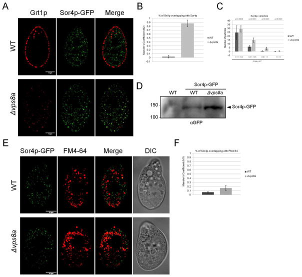
In the endocytic pathway of animals, two related complexes, called CORVET (class C core vacuole/endosome transport) and HOPS (homotypic fusion and protein sorting), act as both tethers and fusion factors for early and late endosomes, respectively. Mutations in CORVET or HOPS lead to trafficking defects and contribute to human disease, including immune dysfunction. HOPS and CORVET are conserved throughout eukaryotes, but remarkably, in the ciliate Tetrahymena thermophila, the HOPS-specific subunits are absent, while CORVET-specific subunits have proliferated. VPS8 (vacuolar protein sorting), a CORVET subunit, expanded to 6 paralogs in Tetrahymena. This expansion correlated with loss of HOPS within a ciliate subgroup, including the Oligohymenophorea, which contains Tetrahymena. As uncovered via forward genetics, a single VPS8 paralog in Tetrahymena (VPS8A) is required to synthesize prominent secretory granules called mucocysts. More specifically, Δvps8a cells fail to deliver a subset of cargo proteins to developing mucocysts, instead accumulating that cargo in vesicles also bearing the mucocyst-sorting receptor Sor4p. Surprisingly, although this transport step relies on CORVET, it does not appear to involve early endosomes. Instead, Vps8a associates with the late endosomal/lysosomal marker Rab7, indicating that target specificity switching occurred in CORVET subunits during the evolution of ciliates. Mucocysts belong to a markedly diverse and understudied class of protist secretory organelles called extrusomes. Our results underscore that biogenesis of mucocysts depends on endolysosomal trafficking, revealing parallels with invasive organelles in apicomplexan parasites and suggesting that a wide array of secretory adaptations in protists, like in animals, depend on mechanisms related to lysosome biogenesis.

Keywords: alveolata; endosomal tether; evolution; extrusome; gene loss; lysosome-related organelle; membrane trafficking; protist.


Conflict of interest statement

DECLARATION OF INTEREST

The authors declare no competing interests.

Figures

Figure 1
Figure 1. Vps8a is essential for mucocyst biogenesis
A) Confocal cross sections, with paired DIC images, of WT, UC616, rescued UC616 and SB281, and Δvps8a. Cells were immunostained with mAbs 5E9 and 4D11, which recognize mucocyst proteins Grl3p (left) and Grt1p (right), respectively. The wildtype pattern of docked mucocysts is absent in UC616 and Δvps8a, but was restored in UC616 and SB281 expressing a VPS8A-GFP transgene. Scale bars, 10μm. B) Electron microscopy. In WT, mucocysts dock at the plasma membrane. In contrast, UC616 and Δvps8a contain cytoplasmic vesicles with granular cores (labeled with asterisks). Scale bars, 500 nm. C) The expression profile of VPS8A (black line) is similar to that of mucocyst-related genes SOR4, STX7L1, APM3, CTH3, CTH4, GRT1, and GRL3. For the plot, based on data downloaded from tfgd.ihb.ac.cn, each value was normalized to that gene’s maximum expression level. The conditions sampled were growing cultures (low Ll, medium Lm, and high Lh culture density), starvation over 24 hours (S0-S24), and timepoints during conjugation (C0-C18). D) Alignment of the VPS8A exon(E2)-intron(I2) junction, and the corresponding translated sequence. The G/A-C/T mutation (black box) results in a premature stop codon (black arrow) 41bp downstream. E) Intron 2 is retained in the VPS8A transcript in UC616. Columns 1 and 2 correspond to WT and UC616 cDNA that was PCR-amplified with BTU1 primers as control; in lanes 3, 4, the primers flanked intron 2 in VPS8A. 2% ethidium bromide-stained gel, with 200 and 300 bp DNA fragments indicated to the left. See also Figure S1.
Figure 2
Figure 2. Multiple mucocyst proteins are not delivered to mucocyst intermediates in Δvps8a cells
A) WT (upper panel) and Δvps8a (bottom panel) cells, co-stained with anti-Grl3p (5E9) and anti-Grt1p (4D11) mAbs conjugated with AlexaFluor 650 and AlexaFluor 488 dyes, respectively. Co-localization between Grt1p and Grl3p is markedly reduced in Δvps8a. Confocal cross sections are shown for clarity. Scale bars, 10μm. B) Percentage of overlap between Grt1p and Grl3p (Mander’s coefficient M1), was calculated with 25 non-overlapping images/sample using Fiji-JACoP plugin. C) Localization of Cth3p-GFP in WT and Δvps8a cells. Cells were immunolabeled with 5E9 anti-Grl3p and rabbit anti-GFP antibodies. Co-localization of Cth3p-GFP with Grl3p is reduced in Δvps8a, and the proteins localize in heterogeneous cytoplasmic vesicles. D) Overlap between Cth3p-GFP and Grl3p was calculated as in (B), based on 40 non-overlapping images/sample. E) Magnification of the boxed area in C (merge), showing co-localization of Cth3p-GFP and Grl3p in a large compartment in Δvps8a. F) Localization of Igr1p-GFP in WT and Δvps8a cells. Cells were immunolabeled with mAb 5E9. Co-localization of Igr1p-GFP with Grl3p is reduced in Δvps8a. G) Overlap between Igr1p-GFP and Grl3p was calculated as in (B), based on 25 non-overlapping images/sample. H) Magnification of the boxed area in F (merge), showing co-localization of Igr1p-GFP and Grl3p in a large compartment in Δvps8a. I and J) CLEM imaging of overexpressed Igr1p-GFP in WT and mutant (SB281, which bears the identical VPS8A mutation as UC616). Fluorescence images (lower images) are paired with the corresponding electron micrographs (upper images). Left, low magnification images. Scale bar, 10μM. The fuschia-stained structures are the Micro- and Macronuclei stained with DAPI; green represents Igr1p-GFP. Right, boxed regions in the left images are shown at high magnification. From top to bottom: EM image, fluorescence image (Igr1p-GFP), fluorescence image inverted (negative to positive) and then overlaid on the electron micrograph to show precise mapping of the fluorescence signals onto cellular structures. In WT, Igr1p-GFP is present in mucocysts. In SB281, Igr1p-GFP does not accumulate in the granular mucocyst-related vesicles (asterisks), but is instead found in electron-dense structures likely to represent degradative compartments. Scale bar, 0.5μm. See also Figure S2.
Figure 3
Figure 3. Model for mucocyst biogenesis in Tetrahymena thermophila
Two different types of vesicles deliver cargo to immature mucocysts. The first is generated directly from a trans Golgi compartment and contains condensed proGrl proteins. These vesicles accumulate in both Δstx7l1 and Δvps8a mutants; similar vesicles, though docked at the plasma membrane, accumulate in Δsor4. The second corresponds to a late endosomal compartment, and transports the protease Cth3p and members of the GRT family, Grt1p and Igr1p, bound to the sorting receptor Sor4p. For simplicity, we depict a single compartment that functions both as a trans Golgi and an early endosome, as has been described in plants[80]; whether these are separate compartments in ciliates is unknown. The CORVET subunit Vps8ap (this paper) and the mucocyst-resident syntaxin Stx7l1p mediate the tethering and fusion between the two classes of vesicles. The AP-3 complex appears required for delivery of some as-yet unidentified mucocyst maturation factors.
Figure 4
Figure 4. Sor4p co-localizes with its ligand Grt1p in Δvps8a but not wildtype cells
A) WT and Δvps8a expressing Sor4p-GFP were immunolabeled with anti-Grt1p mAb. Confocal cross sections are shown for clarity. Scale bars, 10μm. B) Overlap between Grt1p and Sor4p-GFP (M2) was calculated as in (2B), based on 40 non-overlapping images/sample. C) The distribution of Sor4p-GFP vesicle sizes is shifted in Δvps8a compared to wildtype. Shown are Mean particle counts for each size class, calculated using the Fiji tool “Analyze Particles” with 40 non-overlapping images/sample. p-values determined by two-tailed t-test. There are significant increases in vesicle number in Δvps8a compared to WT in multiple size classes: (0.1< >0.2 μm2) = 0.6-fold; (0.2< >0.5 μm2) = 1.6-fold; (0.5< >1 μm2) = 5.8-fold. In addition, rare Sor4p-GFP-positive structures of (1< >3 μm2) appear almost exclusively in Δvps8a. D) SDS-PAGE and Western blotting of whole cell lysates from 1.5×10⁵ WT and Δvps8a cells expressing Sor4p-GFP, and an untransformed WT control, probed with anti-GFP antibody. The position predicted for full-length Sor4-GFP is indicated. Δvps8a cells accumulate more Sor4p-GFP than do WT cells. E) Confocal live microscopy of WT and Δvps8a expressing Sor4p-GFP and pulse labeled with 5 μM FM4-64 for 5min. A small fraction of Sor4p-GFP vesicles in WT cells is labeled with FM4-64, and this overlap increases in Δvps8a. Scale bars, 10 μm. F) Overlap of Sor4p-GFP with FM4-64 (M1) was calculated as in Figure 2B, using 40 non-overlapping images/sample. See also Figure S3.
Figure 5
Figure 5. Expansion in ciliates of CORVET VPS8 genes
A) Expression profiles of the six T. thermophila VPS8 paralogs, displayed as in (1C), shows that each paralog has distinct expression peaks, and only VPS8A (black line) shows the profile associated with mucocyst formation (Figure 1C). Expression data were downloaded from the Tetrahymena Functional Genomics Database (TFGD, http://tfgd.ihb.ac.cn/). B) Non-essential paralogs VPSB, E, and F are dispensable for mucocyst formation. Δvps8b, Δvps8e, and Δvps8f cells were immunolabeled with antibodies against Grl3p or Grt1p, followed by Texas red-conjugated goat anti-mouse. All strains maintained WT patterns of docked mucocysts. Surface and cross sections are indicated at the panel bottom. Scale bars, 10μm. See also Figure S4.
Figure 6
Figure 6. Association of Vps8ap with Rab7 endosomes
A) Distribution of HOPS/CORVET subunits across ciliate diversity. Relationships between ciliate species, based upon [81], are depicted by the evolutionary tree on the left (not to scale), with phylogenetic class indicated in bold. The dotplot on the right depicts the presence/absence of the indicated subunit within each species, as defined in Star*Methods-Method details. Gene IDs are listed in Table S1. B) Cells were transformed to co-express mNeon-tagged Vps8ap at the endogenous locus together with either the Rab5 homolog mCherry-Rab22Ap (upper panel) or mCherry-Rab7p (lower panel), respectively. Rab transgene overexpression was induced with 1 μg/ml CdCl2 for 2.5h in SPP. Scale bars, 10μm. C) The overlap between Vps8ap-mNeon and each Rab-GTPase (M1) was measured as in Figure 2B. Vps8a-mNeon overlapped ~50% with mCherry-Rab7, but only ~12% with mCherry-Rab22A. The Mean M1 values were derived from 65 and 66 non-overlapping images for Rab22Ap and Rab7p samples, respectively. D) and E) Endosome distribution in WT vs Δvps8a cells. Shown are single frames from time-lapse movies of WT and Δvps8a cells, overexpressing mCherry-Rab22Ap (D) and mCherry-Rab7p (E). Cells were transferred to S-medium and mCherry-Rab transgene overexpression was induced as in (B). Rab22Ap-positive endosomes have a similar distribution in WT and Δvps8a, but Rab7p-positive endosomes form large clusters in Δvps8a cells. Scale bars, 10μm. F) Co-immunoisolation of Vps8ap and c-myc-Rabs. Cells were transformed to express Vps8ap-FF-ZZ (flag-ZZ domain) together with either c-myc-Rab22Ap or c-myc-Rab7p, via gene replacement at the endogenous loci. Detergent lysates were incubated with bead-bound anti-c-myc or anti-FLAG Abs, and bead eluates were analyzed by SDS-PAGE and Western blotting using anti-myc or anti-FLAG antibodies. The top panel shows total Vps8a-FF-ZZ in each cell line, while the bottom panel shows total myc-Rab22a or myc-Rab7 in each cell line. The middle panel shows Vps8a-FF-ZZ that was immuno-isolated via interaction with Rab22A (left lane) or Rab7 (right lane). Vps8ap preferentially interacts with Rab7p. See also Figure S5 and S6.

Similar articles

Cited by

References

    1. Palade G. Intracellular aspects of the process of protein synthesis. Science. 1975;189:347–358. - PubMed
    1. Keller P, Simons K. Post-Golgi biosynthetic trafficking. J Cell Sci. 1997;110(Pt 24):3001–3009. - PubMed
    1. Burgoyne RD, Morgan A. Regulated exocytosis. Biochem J. 1993;293:305–316. - PMC - PubMed
    1. Tran DT, Ten Hagen KG. Real-time insights into regulated exocytosis. J Cell Sci. 2017;130:1355–1363. - PMC - PubMed
    1. Orci L, Ravazzola M, Amherdt M, Perrelet A, Powell SK, Quinn DL, Moore HP. The trans-most cisternae of the Golgi complex: a compartment for sorting of secretory and plasma membrane proteins. Cell. 1987;51:1039–1051. - PubMed

Publication types

MeSH terms

Substances

LinkOut - more resources