. 2018 Mar 13;10(3):942-955.
doi: 10.1016/j.stemcr.2018.01.023. Epub 2018 Mar 1.

Exosome-Mediated Benefits of Cell Therapy in Mouse and Human Models of Duchenne Muscular Dystrophy

Affiliations

Exosome-Mediated Benefits of Cell Therapy in Mouse and Human Models of Duchenne Muscular Dystrophy

Mark A Aminzadeh et al. Stem Cell Reports. .

Abstract

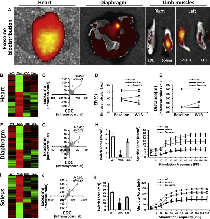
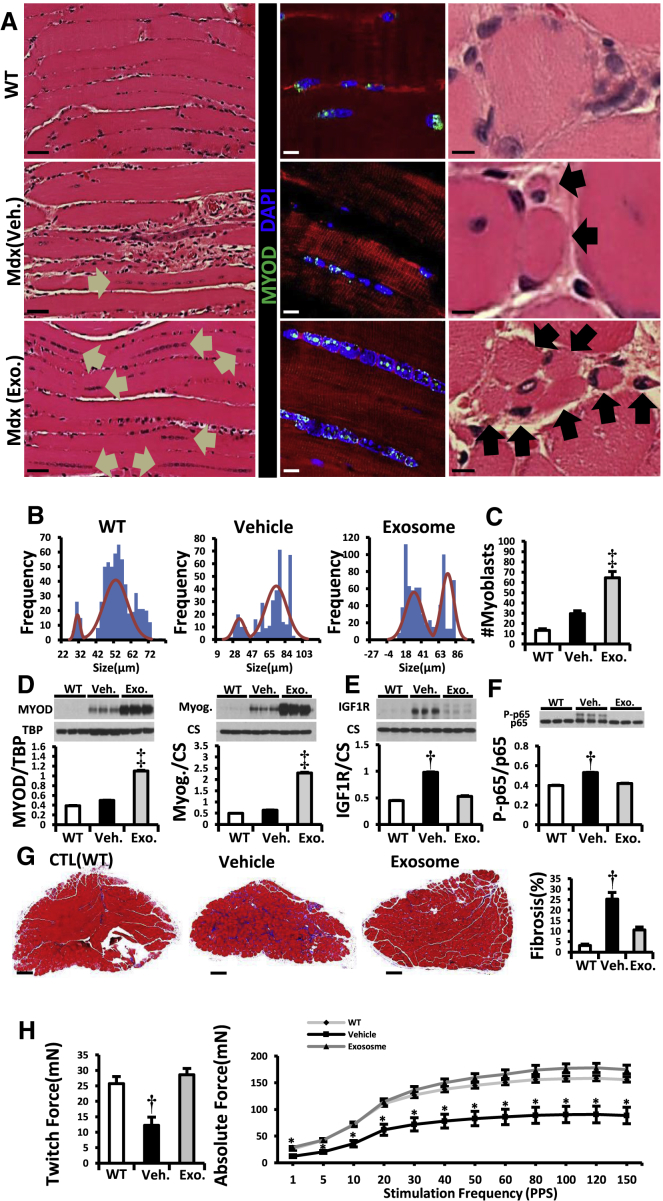
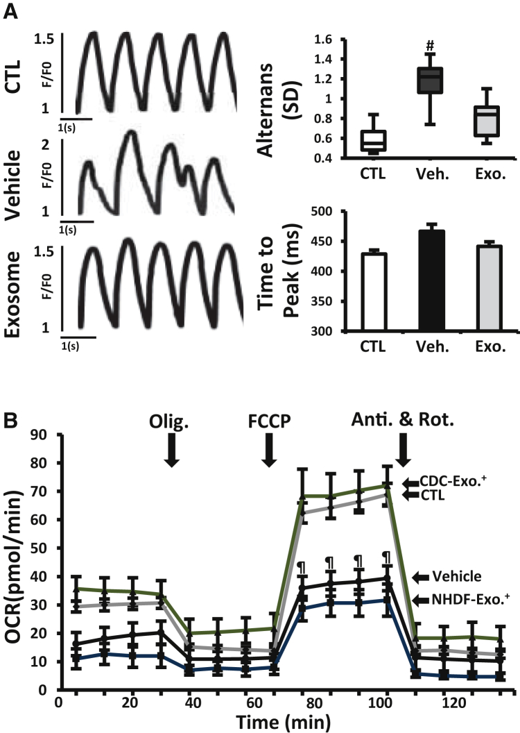
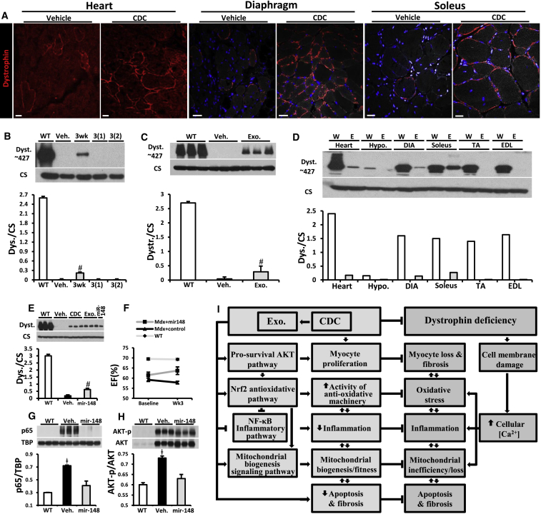
Genetic deficiency of dystrophin leads to disability and premature death in Duchenne muscular dystrophy (DMD), affecting the heart as well as skeletal muscle. Here, we report that clinical-stage cardiac progenitor cells, known as cardiosphere-derived cells (CDCs), improve cardiac and skeletal myopathy in the mdx mouse model of DMD. Injection of CDCs into the hearts of mdx mice augments cardiac function, ambulatory capacity, and survival. Exosomes secreted by human CDCs reproduce the benefits of CDCs in mdx mice and in human induced pluripotent stem cell-derived Duchenne cardiomyocytes. Surprisingly, CDCs and their exosomes also transiently restored partial expression of full-length dystrophin in mdx mice. The findings further motivate the testing of CDCs in Duchenne patients, while identifying exosomes as next-generation therapeutic candidates.

Keywords: cardiomyopathy; cardiosphere-derived cells; dystrophin; exosomes; microRNA; muscular dystrophy.


Figures

None
Graphical abstract
Figure 1
Figure 1
CDC Transplantation into mdx Hearts Function, survival, antioxidant pathways, inflammation, and mitochondrial dysfunction improved by CDC transplantation into mdx mice. (A) Ejection fraction (EF) in CDC-injected mdx mice (Mdx + CDC) and vehicle-injected mdx mice (Mdx + vehicle) in response to injections at baseline (10 months of age) and 3 months later (WT, n = 7; Mdx + vehicle and Mdx + CDC, n = 12 each). (B) Exercise capacity in mice subjected to weekly high-intensity treadmill exercise, starting 3 weeks after single-dose CDC or vehicle administration (WT, n = 7; Mdx + vehicle and Mdx + CDC, n = 11 each). Cardiac (A) and treadmill (B) experiments were performed separately on different groups of experimental mice. (C) Kaplan-Meier analysis of survival in the same animals as (B) shows lower survival in vehicle-treated mdx mice than in CDC-treated mdx mice or WT controls (p < 0.001, log rank test); the latter two groups, however, were statistically comparable. (D) Immunohistochemical images of NRF2 in mdx mouse hearts 3 weeks after administration of vehicle or CDCs. Age-matched WT mice served as control. (E–I) Western blots and pooled data for protein abundance of phospho-AKT (AKT-pT308, AKT-pS473; E), cytoplasmic phospho-NRF2 (NRF2- pS40; F), nuclear NRF2 (G), NRF2 downstream gene product, hemeoxygenase-1 (HO-1; H), and malondialdehyde protein adducts (I) in mdx mouse hearts 3 weeks after administration of vehicle or CDCs (WT, n = 4; Mdx + vehicle and Mdx + CDC, n = 6 each). (J) Immunohistochemical images of hearts stained for inflammatory cell markers CD68, CD20, and CD3. Black arrows point to CD68+ (upper row), CD20+ (middle row), and CD3+ (lower row) cells. (K) Western blots, pooled data, and bar graph (lower right) representing protein abundance of nuclear p65, p-IκB (NF-κB pathway), and MCP1 (monocyte chemoattractant protein1) and average number of indicated inflammatory cells and in mdx mouse hearts. (L) Transmission electron microscopy (TEM) images from mdx mouse hearts 3 weeks after administration of vehicle (Mdx + vehicle) or CDCs (Mdx + CDC). Age-matched WT mice served as control. (M and N) Representative western blots and pooled data for mitochondrial respiratory chain subunits in WT and vehicle/CDC mdx heart tissues (M) and oxygen consumption rate (OCR) of mitochondria isolated from the hearts of WT and CDC- or vehicle-treated mdx mice (N) 3 weeks after treatment (WT, n = 3; Mdx + vehicle and Mdx + CDC, n = 8 each). Substrates (pyruvate, malate, and ADP), a selective uncoupler (FCCP) and blockers (oligomycin [Olig.]; antimycin and rotenone [Anti. & Rot.]) of oxidative phosphorylation were applied when indicated. Pooled data are means ± SEM, CM: cardiomyocyte. p < 0.05 versus Mdx + CDC; #p < 0.005 versus Mdx + CDC; p < 0.05 versus Mdx + Vehicle and WT; p < 0.002 versus Mdx + CDC and WT. Scale bars: 10 μm (D and J); 5 μm (L).
Figure 2
Figure 2
CDC Exosome Injection into mdx Hearts Reproduces the Benefits of CDCs (A) Sustained functional benefit for at least 3 months with each of two sequential CDC exosome injections in mdx mice (n = 11). (B) Western blots and pooled data for cardiac collagen IA and IIIA. (C) Immunohistochemical images and pooled data (WT, n = 4; vehicle and CDC exosome-treated [Mdx (XO)], n = 6 each) from mdx mouse hearts stained for Ki67 [upper row] and Aurora B [lower row]. Arrows point to Ki67+ (upper row) and Aurora B+ (lower row) cardiomyocytes. Data are means ±SEM; p < 0.05 versus Mdx + exosome; p < 0.01 versus Mdx + exosome and WT; p < 0.02 versus Mdx + vehicle and WT mice. Scale bar: 10 μm.
Figure 3
Figure 3
CDC Transplantation in mdx Hearts Conferred Beneficial Effects on Diaphragm and Soleus Muscles (A) Two-dimensional hierarchical clustering using genes with at least two times fold change difference between vehicle/CDC mdx diaphragms. (B and C) Western blots and pooled data for protein abundance of malondialdehyde protein adducts (B), cytoplasmic p-p65 and p-IκB (C; NF-κB pathway; WT, n = 4; vehicle and CDC, n = 6 each) and immunohistochemical images of diaphragm stained for inflammatory cell markers CD20 and CD3; bar graph represents the average number of indicated inflammatory cells 3 weeks after administration of vehicle or CDCs into mdx hearts. (D) Representative Masson trichrome images and morphometric analysis in diaphragms 3 weeks after administration of vehicle or CDCs into the hearts of mdx mice. (E–G) In vitro measurement of isometric diaphragm contractile properties: twitch force (E), maximum tetanic force (F), and force/frequency relationships (G) 3 weeks after CDC/vehicle mdx heart treatments. (H) Two-dimensional hierarchical clustering using genes with at least two times fold change difference between vehicle/CDC mdx soleus. (I–K) In vitro measurement of isometric soleus contractile properties: twitch force (I), maximum tetanic force (J), and force/frequency relationships (K) 3 weeks after CDC/vehicle treatment of mdx hearts. (L) Correlation of fold changes in expression of same genes in diaphragm and soleus 3 weeks after intramyocardial CDC injection in mdx mice. (M) Three-dimensional plot depicting principal components analysis (PCA) of RNA-seq expression data from exosomes isolated from hypoxic conditioned media and effluents of CDC- or vehicle-treated mdx hearts. The effluent of isolated mdx hearts undergoing Langendorff perfusion was collected for exosome isolation and subsequent RNA-seq 3 days after intramyocardial CDC/vehicle injection. PCA analysis showed clustering of CDC exosomes (red) with exosomes isolated from effluent of CDC mdx hearts (blue), but not vehicle-injected mdx hearts (stippled), indicating that CDC exosomes were shed from mdx hearts at least 3 days after intramyocardial CDC injection. Effluents of mdx hearts from the same group were pooled (n = 3 for each group). Data are means ±SEM; p < 0.05 versus Mdx + CDC; p < 0.05 versus Mdx + CDC and WT mice. Scale bars: 10 µm (C); 100 µm (D).
Figure 4
Figure 4
Systemic CDC Exosome Injection Mimicked the Cardiac and the Remote Effects of Intramyocardial CDC Injection in mdx Mice (A) Systemic biodistribution of CDC exosomes after intraventricular injection in mdx mice. CDC exosomes were stained with fluorescent lipid dye and tracked 6 hr later using bioluminescence imaging. (B) Two-dimensional hierarchical clustering using genes from hearts of non-treated mdx mice and of mdx mice treated intramyocardially with CDCs or intraventricularly with CDC exosomes. Genes with at least 2-fold differences with corresponding transcripts in non-treated mdx mice were included. (C) Correlation of fold changes in expression of same genes 3 weeks after intramyocardial CDC injection or intraventricular CDC exosome injection in mdx hearts. (D and E) EF and exercise capacity in mdx mice 3 weeks after intraventricular injection of vehicle/CDC exosome (WT, n = 5; Mdx + vehicle and Mdx + CDC exosome, n = 9 each). (F) Two-dimensional hierarchical clustering using genes from diaphragm of non-treated mdx mice and of mdx mice treated intramyocardially with CDCs or intraventricularly with CDC exosomes. Genes with at least 2-fold differences with corresponding genes in non-treated mdx mice were included. (G) Correlation of fold changes in expression of the same genes in diaphragm 3 weeks after intramyocardial CDC injection or intraventricular CDC exosomes injection. (H) Diaphragm isometric twitch force and force/frequency relationships 3 weeks after intraventricular CDC exosome injection. (I–K) Two-dimensional hierarchical clustering (I), correlation analysis (J), and isometric twitch force and force/frequency relationships (K) from soleus muscle 3 weeks after intraventricular CDC exosome injection. Data are means ± SEM; p < 0.05 versus Mdx + CDC exosome; p < 0.05 versus Mdx + CDC exosome and WT mice.
Figure 5
Figure 5
Intramuscular Injection of CDC Exosomes Resulted in Muscle Growth and Reversal of Pathophysiologic Abnormalities (A) H&E and immunohistochemical images of soleus muscle stained for MYOD (WT, vehicle, and CDC exosome-treated [Mdx (exosome)] mdx mouse soleus). Arrows in H&E images point to the lined-up nuclei (left column) and myofibers (right column). In the immunohistochemistry, linearly arranged nuclei were positive for MYOD (middle column). (B and C) Frequency distribution of myofiber sizes and number of myoblasts (MYOD+) 3 weeks after vehicle/CDC exosome injection in mdx soleus muscles (WT, n = 5; vehicle and exosome, n = 9 each). (D–F) Western blots and pooled data for protein abundance of MYOD, myogenin (D), IGF1 receptor (IGF1R; E) and cytoplasmic p-p65 (F) in mdx soleus muscles 3 weeks after intrasoleus vehicle/CDC exosome injection (WT, n = 4; vehicle and exosome, n = 6 each). (G) Representative Masson trichrome images and morphometric analysis in mdx soleus muscles 3 weeks after administration of vehicle or CDC exosomes into mdx soleus (WT, n = 5; vehicle and exosome. n = 9 each). (H) In vitro measurement of soleus isometric twitch force and force/frequency relationships 3 weeks after vehicle/CDC exosome injection into mdx soleus muscles. Pooled data are means ± SEM. p < 0.05 versus Mdx + CDC exosome; p < 0.05 versus Mdx + CDC exosome and WT mice; p < 0.002 versus Mdx + vehicle and WT mice. Scale bars: 5 μm (A, right column), 10 μm (A, middle column), 50 μm (A, left column), and 200 μm (G).
Figure 6
Figure 6
CDC Exosomes in Human Duchenne Cardiomyocytes Derived from iPSCs (A) Calcium transients from normal and DMD CM measured during 1 Hz burst pacing. Duchenne cardiomyocytes were primed with vehicle or CDC exosomes (exosomes) 1 week before assessment. Bar graphs of calcium transient alternans (variation in beat-to beat calcium transient amplitude) and time to peak (n = 10 cells in each group). (B) Oxygen consumption rate (OCR) in DMD CMs primed with CDC exosomes or exosomes from normal human dermal fibroblasts (NHDF, as control; NHDF exosome) 1 week before OCR measurement. Normal (CTL) and non-treated DMD CM (vehicle) were studied in parallel. Results from four independent experiments performed in three replicates are shown. See Figure 1 legend for abbreviations. All data are means ± SEM except for the boxplot (means ± SD). #p < 0.03 versus CDC exosome and CTL (normal cardiomyocyte); p < 0.02 versus CDC exosome.
Figure 7
Figure 7
Exosomes Mediate Reversal of Key Pathophysiological Features of Duchenne Muscular Dystrophy (A) Immunohistochemical images of dystrophin in mdx mouse heart, diaphragm, and soleus treated with and without CDC at 10 months of age. (B) Western blot and pooled data for dystrophin protein in WT control mouse heart and mdx mouse hearts 3 weeks and 3 months after first intramyocardial CDC injection, 3(1), and 3 months after second (repeat) CDC injection into myocardium, 3(2), versus Mdx + vehicle (Veh.); (Mdx + vehicle and Mdx + CDC, n = 6 each). (C) Western blot and pooled data for dystrophin protein in WT control mouse heart and mdx mouse hearts 3 weeks after CDC exosome injection into myocardium (Mdx + vehicle and Mdx + exo, n = 6 each). (D) Western blot showing protein content of dystrophin in WT control and in mdx mouse (Exo) heart, hypothalamus (hypo.), diaphragm (DIA), soleus, tibialis anterior (TA), and extensor digitorum longus (EDL) 1 week after systemic CDC exosome delivery by intraventricular injection (n = 2). (E) Western blots and pooled data for protein abundance of dystrophin isoform: dp427 in mdx mouse hearts 3 weeks after intramyocardial injection of vehicle, CDC, CDC exosomes, or mimics of Mir-148a. (F) Ejection fraction (EF) at baseline and 3 weeks after intramyocardial injection of Mir-148a or miRNA control in mdx mice. WT EF values are also shown for reference, n = 5 per group. (G and H) Western blots and pooled data for nuclear p65 (G) and phosphorylated AKT (H) in mdx mouse hearts 3 weeks after Mir-148a treatment (vehicle and Mir-148a, n = 6 each). (I) Schematic of pathophysiological mechanisms operative in Duchenne cardiomyopathy and the cellular mechanisms recruited by CDCs and their exosomes. All organs were from mice 10 months old at baseline. All data are means ± SEM. #p < 0.05 versus Mdx + vehicle and WT; ǂ p < 0.05 versus Mir-148a and WT. Scale bars: 25 μm (A, heart); 25 μm (A, diaphragm); 20 μm (A, soleus).

References

    1. Advani V.M., Dinman J.D. Reprogramming the genetic code: the emerging role of ribosomal frameshifting in regulating cellular gene expression. Bioessays. 2016;38:21–26. - PMC - PubMed
    1. Aminzadeh M.A., Tobin R., Smith R., Marban L., Marban E. Heart-derived cell therapy for Duchenne cardiomyopathy: cardiosphere-derived cells and their exosomes improve function, restore mitochondrial integrity and reverse degenerative changes in the hearts of Mdx mice. Circ. Res. 2014;115:e90.
    1. Aminzadeh M.A., Durvasula P., Tobin R., Guan X., Andres A., Taylor D., Ibrahim A., Sun B., Torrente A., Goldhaber J. Exosome-mediated reversal of Duchenne cardiomyopathy. Circulation. 2015;132:A16015.
    1. Aminzadeh M.A., Tseliou E., Sun B., Cheng K., Malliaras K., Makkar R.R., Marbán E. Therapeutic efficacy of cardiosphere-derived cells in a transgenic mouse model of non-ischaemic dilated cardiomyopathy. Eur. Heart J. 2015;36:751–762. - PMC - PubMed
    1. Ascheim D., Jefferies J.L. 2016. A randomized, open-label study of the safety and efficacy of multi-vessel intracoronary delivery of allogeneic cardiosphere-derived cells in patients with cardiomyopathy secondary to Duchenne muscular dystrophy.https://clinicaltrials.gov/ct2/show/NCT02485938

Publication types

MeSH terms