. 2018 Sep;32(9):1958-1969.
doi: 10.1038/s41375-018-0012-5. Epub 2018 Jan 31.

Distinct roles for phosphoinositide 3-kinases γ and δ in malignant B cell migration

Affiliations

Distinct roles for phosphoinositide 3-kinases γ and δ in malignant B cell migration

Ahmed Y Ali et al. Leukemia. 2018 Sep.

Abstract

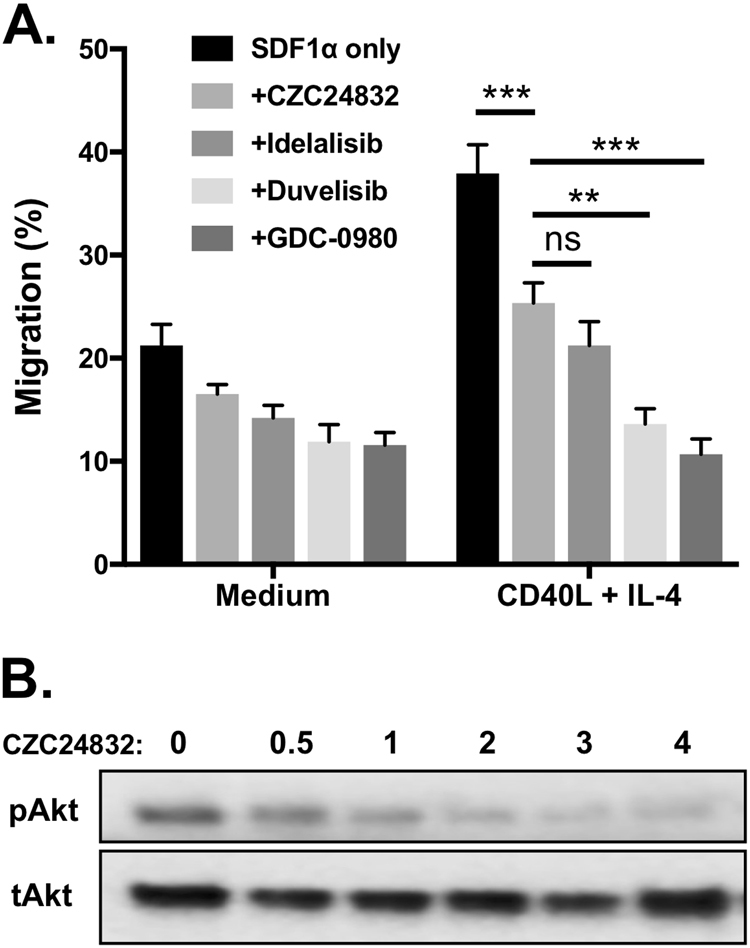
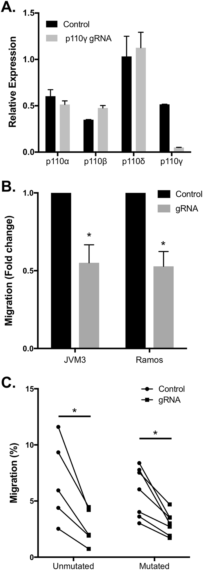
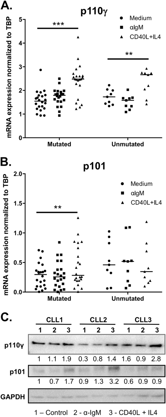
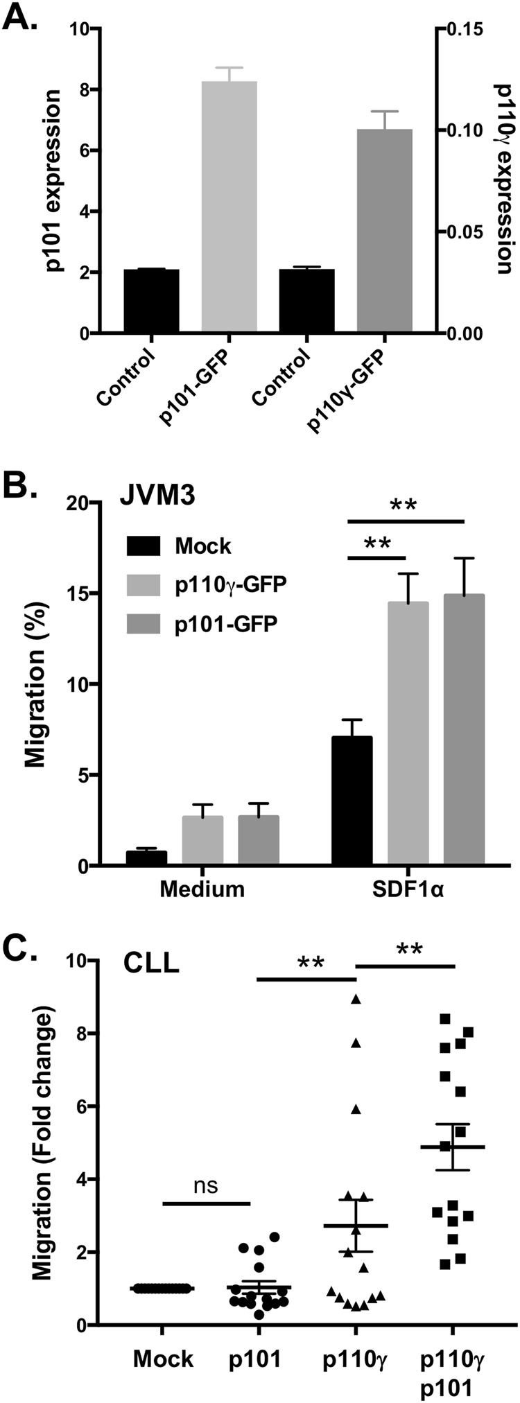
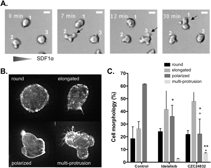
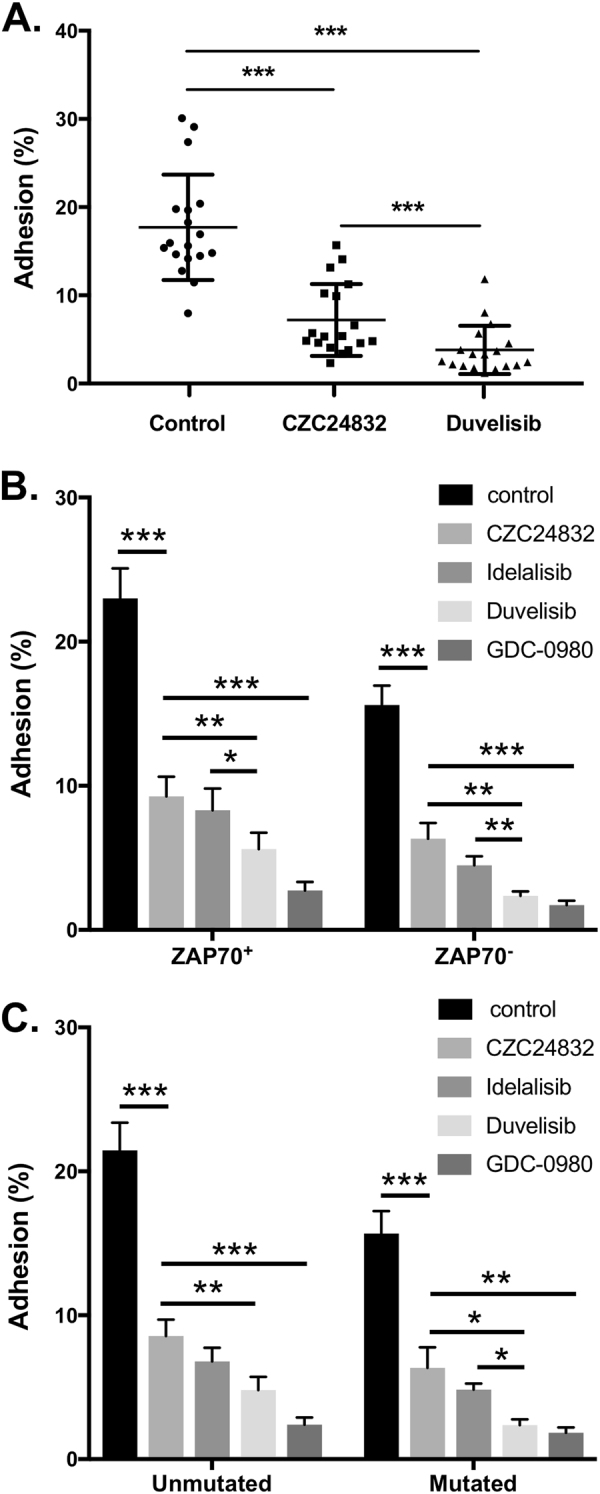
The PI 3-kinases (PI3K) are essential mediators of chemokine receptor signaling necessary for migration of chronic lymphocytic leukemia (CLL) cells and their interaction with tissue-resident stromal cells. While the PI3Kδ-specific inhibitor idelalisib shows efficacy in treatment of CLL and other B cell malignancies, the function of PI3Kγ has not been extensively studied in B cells. Here, we assess whether PI3Kγ has non-redundant functions in CLL migration and adhesion to stromal cells. We observed that pharmaceutical PI3Kγ inhibition with CZC24832 significantly impaired CLL cell migration, while dual PI3Kδ/γ inhibitor duvelisib had a greater impact than single isoform-selective inhibitors. Knockdown of PI3Kγ reduced migration of CLL cells and cell lines. Expression of the PI3Kγ subunits increased in CLL cells in response to CD40L/IL-4, whereas BCR cross-linking had no effect. Overexpression of PI3Kγ subunits enhanced cell migration in response to SDF1α/CXCL12, with the strongest effect observed within ZAP70 + CLL samples. Microscopic tracking of cell migration within chemokine gradients revealed that PI3Kγ functions in gradient sensing and impacts cell morphology and F-actin polarization. PI3Kγ inhibition also reduced CLL adhesion to stromal cells to a similar extent as idelalisib. These findings provide the first evidence that PI3Kγ has unique functions in malignant B cells.


Conflict of interest statement

The authors declare that they have no conflict of interest.

Figures

Fig. 1
Fig. 1
Inhibition of PI3Kγ impairs Akt phosphorylation and B cell migration in a transwell assay. a Inhibition of PI3Kγ or PI3Kδ significantly decreased the migration of JVM3 cells. JVM3 cells were cultured in medium or stimulated for 24 h with CD40L/IL-4 and then incubated with the PI3Kγ-specific inhibitor CZC24832 (2 µM), the PI3Kδ-specific inhibitor idelalisib (1 µM), dual PI3Kδ/γ inhibitor duvelisib (1 µM), or the pan-PI3K inhibitor GDC0980 (1 µM) and subjected to a transwell migration assay. DMSO served as the vehicle control for the inhibitors, while SDF1α (100 ng/ml) served as the chemoattractant (n = 7). b Effect of PI3Kγ inhibitor on Akt phosphorylation. JVM3 cells were pre-incubated with the indicated concentrations of CZC4832 (in μM) and then stimulated with SDF1α for 10 min. Akt Ser473 phosphorylation and total Akt levels were assessed by Western blot
Fig. 2
Fig. 2
Inhibition of PI3Kγ or PI3Kδ impairs CLL cell migration. a Effect of PI3Kγ inhibitor on CLL cell migration. CLL cells were cultured in medium or stimulated with CD40L+IL-4 for 24 h and subjected to a transwell migration assay. Graphs represent migration of cells from individual CLL patient samples with or without addition of PI3Kγ inhibitor CZC24832, connected by lines. b Inhibition of PI3Kγ or PI3Kδ decreases the migration of CLL cells to a similar extent, while dual PI3Kδ/γ inhibition using duvelisib has significantly greater effect than PI3Kγ inhibitor alone. c Combination of PI3Kγ inhibitor and PI3Kδ inhibitor decreases the migration of CLL cells to a greater extent than either inhibitor alone. Lines connect individual patient sample migration responses in the presence of the indicated inhibitors. Note all inhibitor-treated groups were significantly different than the control untreated group, whereas the CZC24832+idelalisib combination was not significantly different than duvelisib.
Fig. 3
Fig. 3
The effect of CRISPRi knockdown of p110γ on the migration of malignant B cell lines and CLL cells. a Confirmation of p110γ CRISPRi knockdown specificity. JVM3 cells were transfected with either dCas9-GFP expression vector alone (Control) or co-transfected with dCas9-GFP plus vectors expressing p110γ-targeting guide RNAs (at a 1:3 ratio). After 24 h, p110 isoform expression within sorted GFP+ transfectants was assessed by qPCR. b p110γ knockdown significantly impaired the migration of JVM3 and Ramos cells. Cells were transfected as above and then subjected to a transwell migration assay. Data represent average fold change in migration of GFP+ cells (n = 3). c p110γ knockdown significantly reduced the migration of primary CLL cells regardless of their IgVH mutation status. CLL cells were transfected with either dCas-GFP expression vector or dCas9-GFP plus vectors expressing p110γ-targeting guide RNAs then assessed 48 h later using a transwell migration assay. Percent of cells migrating toward SDF1α was determined by flow cytometry counting of live GFP-expressing cells present in the upper and lower chambers after 3 h
Fig. 4
Fig. 4
Expression of PI3Kγ subunits in malignant B cell lines and CLL patient samples. a, b CLL cells were stimulated with F(ab′)2 α-IgM (10 µg/ml) or CD40L/IL-4 (50 ng/ml each) and harvested 24 h later for RNA extraction and RT-qPCR analysis. mRNA expression of a p110γ and b p101 were determined, and expression levels were normalized against the expression of TATA box-binding protein (TBP). Patients were divided into indolent vs. progressive groups based on IgVH mutation status. c Protein expression of p110γ and p101 in CLL samples in response to BCR stimulation or CD40L/IL-4 stimulation. Data are representative of nine patients analyzed
Fig. 5
Fig. 5
Overexpression of PI3Kγ subunits affects the migration of malignant B cells. a Confirmation of p110γ-GFP and p101-GFP overexpression in JVM3 cells. JVM3 cells were transfected with either p101-GFP or p110γ-GFP plasmids. 24 h post transfection, RNA was harvested and expression of p101 or p110γ was determined by RT-qPCR analysis. b Overexpression of p110γ or p101 enhances the migration of JVM3 in response to the chemokine SDF1α. 24 h after transfection with p101-GFP plasmid or p110γ-GFP, migration of live GFP-expressing cells in response to SDF1α (100 ng/ml) was assessed using a transwell migration assay. The graph shows the mean and SEM of four experiments. c Overexpression of PI3Kγ subunits enhances CLL cell migration. p110γ-GFP, p101-GFP, or both together were overexpressed in CLL cells. After 48 h, migration of live GFP+ cells was assessed by transwell assays as above. Results are expressed as fold change in migration relative to the control transfection of the same patient sample
Fig. 6
Fig. 6
Impact of PI3Kγ or PI3Kδ inhibition on directional migration behavior. JVM3 cells were seeded into a collagen gel and treated with the indicated inhibitors for 60 min. After establishing the SDF1α gradient, time-lapse imaging was performed and cell migration tracks were analyzed using the IMARIS 8.0 software. a Plots illustrate cell tracks from a single representative experiment, overlaid to the same origin. Note that cells treated with CZC24843 show visibly impaired chemotaxis toward the higher SDF1α concentration (right half of the graphs). bc Quantitative analysis of cell tracks showing that both PI3Kδ and PI3Kγ inhibition reduced the chemotactic index b and migration velocity c. Mann–Whitney test, **p < 0.01; ***p < 0.001
Fig. 7
Fig. 7
PI3Kδ and PI3Kγ inhibitors differentially affect cytoskeletal remodeling and cell morphology. a Time-lapse images of CZC24832-treated Ramos cells migrating within SDF1α gradient, showing the dynamic formation and retraction of multiple protrusions and failure to form a stable polarized morphology. Note that all three cells in this field exhibit multiple protrusions at one of the time points (indicated by arrows) and failed to migrate significantly in the direction of the SDF1α gradient. b Morphological scoring demonstrating the differential impact of PI3Kδ and PI3Kγ inhibitors on Ramos cell polarization. Ramos cells were pretreated with inhibitors, stimulated with 100 ng/ml SDF1α for 1 min, then fixed and F-actin stained using Alexa-488 phalloidin. Representative CZC24832-treated cells exhibiting the four major observed morphologies are shown. c Frequency of cells exhibiting each morphology within control and inhibitor-treated groups. Data are based on three independent experiments scoring over 200 cells per treatment group in total. Paired t-test, asterisk denotes significance comparing drug-treated to control, plus denotes significance comparing CZC24832 to Idelalisib
Fig. 8
Fig. 8
Inhibition of either PI3Kγ or PI3Kδ significantly decreases the adhesion of CLL cells to stromal cells without affecting CLL cell survival. CLL cells were incubated for 24 h with established monolayers of human stromal cell line HS-5 in the presence of the indicated inhibitors. The adhered and non-adhered CLL cell fractions were counted by flow cytometry, gating on the live cell population expressing CD19, to determine the percent adhesion. a Impact of PI3Kγ or PI3Kγ/PI3Kδ dual inhibitors on stromal cell binding. The graph displays percent adhesion of individual CLL patient samples under different inhibitor treatment conditions. Inhibition of PI3Kγ or PI3Kδ significantly decreased the adhesion of CLL cells regardless of b ZAP70 status or c IgVH mutation status

References

    1. Stevenson FK, Krysov S, Davies AJ, Steele AJ, Packham G. B-cell receptor signaling in chronic lymphocytic leukemia. Blood. 2011;118:4313–20. doi: 10.1182/blood-2011-06-338855. - DOI - PubMed
    1. Burger JA, Gribben JG. The microenvironment in chronic lymphocytic leukemia (CLL) and other B cell malignancies: insight into disease biology and new targeted therapies. Semin Cancer Biol. 2014;24:71–81. doi: 10.1016/j.semcancer.2013.08.011. - DOI - PubMed
    1. Lagneaux L, Delforge A, Bron D, De Bruyn C, Stryckmans P. Chronic lymphocytic leukemic B cells but not normal B cells are rescued from apoptosis by contact with normal bone marrow stromal cells. Blood. 1998;91:2387–96. - PubMed
    1. Munk Pedersen I, Reed J. Microenvironmental interactions and survival of CLL B-cells. Leuk Lymphoma. 2004;45:2365–72. doi: 10.1080/10428190412331272703. - DOI - PubMed
    1. Burger JA, Ghia P, Rosenwald A, Caligaris-Cappio F. The microenvironment in mature B-cell malignancies: a target for new treatment strategies. Blood. 2009;114:3367–75. doi: 10.1182/blood-2009-06-225326. - DOI - PMC - PubMed

Publication types

MeSH terms